Deep neck abscess
I Made Nudi Arthana
Deep neck abscess is defined as a localized collection of pus that forms in a potential space between the deep neck fascia due to tissue damage that is the spread of infection from various sources, such as teeth, mouth, throat, paranasal sinuses, middle ear and neck.
LITERATUR REVIEW
Anatomy
Knowledge of the spaces in the neck and their relationship to the fascia is important for diagnosing and treating neck infections. The spaces formed by various fascia on this neck is is potential areas for the occurrence infection. invasion from bacteria will produce cellulitis or abscess, and spread through various routes including through the lymphatic channels. 2
The submandibular space is located between the mucosa of the floor of the mouth (as the superior boundary) and the superficial layer of the deep cervical fascia (as the inferior boundary). Inferiorly, it is formed by the digastric muscle. Laterally, its boundaries are skin, muscle platysma, And corpus mandible. While in the medially it is bordered by the hyoglossus and mylohyoid. Anteriorly, it is bordered by the anterior digastric muscle and the mylohyoid. Posteriorly it is bordered by the submandibular ligament and the posterior digastric muscle. 2
The submandibular space is the space above the hyoid which consists of the sublingual space and the submaxillary space. The sublingual space is separated from the submaxillary space by the mylohyoid muscle. The submaxillary space is further divided into the submental space And room submaxillary (lateral) by muscle digastric front but These two spaces communicate freely. However, there is another division that does not include the sublingual space into the submandibular space, and divides the submandibular space into the submental space and the submaxillary space only. 2,3
The sublingual space contains the sublingual gland, Wharton's duct, and the hypoglossal nerve. It is located above the mylohyoid muscle but still anterior to the tongue, and lateral to the intrinsic muscles of the tongue (genioglossus and geniohyoid) and superior and medial to the mylohyoid muscle. Anteriorly, it borders along the genu of the mandible and posteriorly communicates freely with the submaxillary space. 4
The submaxillary space is beneath the mylohyoid muscle, and contains glands submandibular And gland sap clear. Room submksila This communicates freely with the sublingual space along the posterior edge of the mylohyoid muscle. The submandibular gland is located between the two spaces. 4,5
The submental space is a triangular space located in the midline below the mandible where the superior and lateral borders are limited by the anterior part of the digastric muscle. The base of this space is the mylohyoid muscle while the roof is skin, superficial fascia, platysma muscle. The submental space contains several lymph nodes and fibrous fat tissue. 2,4
The submandibular space is bounded by the oral mucosa of the floor of the mouth superiorly. And restricted by layer superficial fascia cervical in in part inferior space extending from the mandible to the hyoid bone. The submandibular space consists of the sublingual space and the submaxillary space, both of which are separated by the myohyoid muscle. The submaxillary space is further divided into the submental space and the submaxillary (lateral) space by the anterior digastric muscle. The mylohyoid muscle divides submandibular space become room sublingual (superior) Which containing gland sublingual, a small portion of the submandibular gland, Wharton's duct, and the submaxillary (inferior) space containing lymph nodes. The mandibular space is bounded laterally by the inferior line of the body of the mandible, medially by the anterior belly of the digastric muscle, posteriorly by the stylohyoid ligament and the posterior belly of the digastric muscle, superiorly by the mylohyoid and hyoglossus muscles, and inferiorly by the superficial from deep cervical fascia. Room This contain glandular sub mandibular saliva and sub mandibular lymphanodes. 6
However, there are other divisions that do not include the submandibular space and share room submandibular on room submental And room submaxillary only. Abscess can formed in room submandibular or Wrong One its components as a continuation of infection from the head and neck area. 2
The submandibular space communicates with several nearby structures, therefore a submandibular abscess can spread to nearby structures.

Picture 1. Fascia Cervical 14
On area neck there is a number of room potential Which restricted by Cervical fascia. The cervical fascia consists of layers of fibrous connective tissue that enclose organs, muscles, nerves and blood vessels and divide the neck into several potential spaces. The cervical fascia is divided into two parts, namely the superficial cervical fascia and the deep cervical fascia. 4
Fascia cervical superficial located appropriate under skin neck walk from its attachment in process zygomaticus on part superior And walk to down towards the thorax and axilla which consists of subcutaneous fat tissue. The space between the superficial cervical fascia and the deep cervical fascia contains superficial lymph nodes, nerves and blood vessels including the external jugular vein. 4
Fascia cervical deep consists of from three layers namely: 4
Layer superficial
This layer completely envelops the neck, starting from the base of the skull. until area thorax And axillary. On part front spread to area face and attached to clavicle and envelops the sternocleidomastoid muscle, muscle trapezius, masseter muscle, gland parotid and submaxillary. This layer is also called the external layer, investing layer, covering layer and anterior layer.
Layer media
Layer This shared on two division that is division muscular and viscera. Muscular division located under layer superficial fascia cervical deep and wraps the sternohyoid muscle, sternothyroid muscle, thyrohyoid muscle and omohyoid muscle. In the superior part it is attached to the hyoid bone and thyroid cartilage and in the inferior part it is attached to the sternum, clavicle and scapula. The viscera division wraps the anterior organs of the neck that is gland thyroid, trachea And esophagus. In adjacent The posterosuperior layer starts from the posterior part of the skull base to the esophagus while the anterosuperior part is attached to the thyroid cartilage and the hyoid bone. This layer runs down to the thorax, covering the trachea and esophagus and uniting with the pericardium. The buccopharyngeal fascia is part from viscera division is at in the section posterior pharynx and covers the constrictor and buccinator muscles.
Layer deep
This layer is divided into two divisions, namely the alar and prevertebral divisions. The alar division is located between the medial layer of the deep cervical fascia and the prevertebral division, which runs from the base of the skull to the thoracic vertebrae. II and unites with the visceral division of the medial layer of the deep cervical fascia. The alar division completes the posterolateral part of the retropharyngeal space and forms the anterior wall of the danger space. The prevertebral division lies anterior to the vertebral bodies and extends laterally to the transverse processes and covers the muscles in that area. It runs from the base of the skull to the coccyx and forms the posterior wall of the danger space and the anterior wall of the vertebral bodies. These three layers of the deep cervical fascia form the carotid sheath which runs from the base of the skull through the pharyngomaxillary space to the thorax. Room potential neck in shared become room Which involving areas along the neck, suprahyoid space and infrahyoid space. 4
Room Which involving throughout neck consists of from: 4
room retropharyngeal
room danger ( danger space )
room prevertebral
Room suprahyoid consists of from:
room submandibular
room parapharyngeal
room parotid
room mastication
room peritonsil
room temporalis
Room :
A room pretracheal
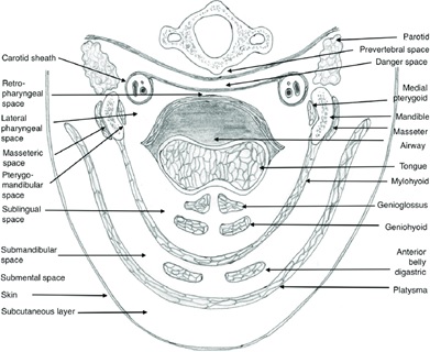
Picture 2 . Potential Space Neck In 1
Epidemiology
In 1997 to 2002, there were 185 cases of deep neck infection. Submandibular abscess (15.7%) was the second most common case after parapharyngeal abscess (38.4), followed by Ludwig's angina (12.4%), parotid (7%) and retropharyngeal (5.9%). 5
On 100 case abscess neck in Which investigated April 2001 until October 2006, the ratio between men and women was 3:2. Submandibular abscess was the most common case (35%), followed by parapharyngeal abscess (20%), masticator (13%), peritonsil (9%), sublingual (7%), parotid (3%), infra
hyoid (26%), retropharyngeal (13%), room carotid (11%). 6.7
Infection neck in donate around 3,400 take care stay every year in the United States. A study using the Kids' Inpatient Database (KID) For determine incident infection neck in find that abscess event retropharyngeal increase in a way significant from 0.1/10,000 on year 2000 to 0.22/10,000 on year 2009. Incident peritonsillar abscess in the year 2009 was 0.94 cases/10,000, and parapharyngeal abscess was 0.14 cases/10,000. 7,16,21
Brito et al reported a total of 16 cases of deep neck abscess in Brazil in 2017. Deep neck abscess is more serious in adults than in children due to the involvement of multiple spaces and causing more complications. Mortality varies. from 1.6% until 7.6% with burden cost system health. 50-70% of cases originate from odontogenic infections although they can be caused by other causes such as upper respiratory tract infections, trauma, parotitis, foreign bodies in the respiratory tract, history of instrumentation and intravenous drug application. The cause is unknown in 8-57% of cases. 21
A study epidemiology abscess neck in on House Sick tertiary in Mexico on 2021 show prevalence 42 case in 5 year, incident 8.4 per year. The mean age of patients was 45.2 years and there was no gender difference. Majority case about two or more from 3 room. More from Half of the cases underwent surgical drainage. The most common comorbidity was type 2 diabetes mellitus. 21
Incident abscess brain is 1 per 100,000 population. Usually arise after trauma or surgery (10%-20% case) or secondary consequence focus septic (20%- 30% case) or spread through expansion direct or track hematology (20%- 40% of cases). 17
Etiology
Most abscesses are caused by multiple microbes, for example they contain mixed flora, and studies have shown that more than 5 species can be isolated from a single case. 1
On room submandibular, infection can be sourced from tooth, floor of the mouth, pharynx, tonsils, sinuses, and salivary glands or submandibular lymph nodes. It may also be a continuation of other deep neck space infections. The causative germs are usually a mixture of aerobic and anaerobic germs. 1.5
Bacterial proliferation and bacterial invasion through enamel organ causes bone necrosis around the tooth root. This usually occurs in patients undergoing dental treatment and drainage of tooth root abscesses. If the tooth root abscess is not drained and not checked, the infection can spread with the abscess to the neck and mediastinum. Infection mostly spread from tooth mandible. And in some case from oral mucosal wounds. Abscess can also due to by trauma, tonsillitis tongue or salivary gland disease. Infection can spread to room deep neck, to the submandibular space, parapharyngeal space and retropharyngeal space. The prevertebral space may also be involved. Deep neck space infections can cause different complications that can be life-threatening such as obstruction upper respiratory tract and mediastinitis. And when all three submandibular spaces (bilateral submandibular and sublingual spaces) are infected, it is called Ludwig's angina. 6
Infection can originate from teeth, floor of mouth, pharynx, submandibular lymph nodes. It is also possible that infection continues from other deep neck spaces. As many as 61% of submandibular abscess cases are caused by dental infections. 7
Infection in this space originates from the second and third molar teeth of the mandible, if the apex is found in lower adhesion from mylohyoid muscle. infection from the teeth can spread to the submandibular space through several routes, namely directly through myolohyoid border, posterior to room sublingual, periostitis And through room masticor. 3
Most deep neck abscesses are caused by a mixture of different germs, Good germs aerobic, anaerobic, and also optional anaerobic. Germs aerobic which are often found are Staphylococcus, Streptococcus sp, Haemophilus influenza, Streptococcus Pneumonia, Moraxtella catarrhalis, Klebsiella sp, Neisseria sp. Anaerobic germs which are often found in deep neck abscesses are gram-negative rod groups, such as Bacteroides, Prevotella, and Fusobacterium. 6
Deep neck infections have the potential to occur in the pharyngeal space. The source of infection can come from from tooth , pharynx, or due to trauma to channel breath on , where there is perforation in the protective mucous membrane of the mouth or pharynx. 7
Besides That, infection gland saliva, infection channel breath on, object foreign and medical device intervention ( iatrogenic ) can be a factor causing deep neck abscess. However, there are still about 20% of cases that occur, the cause is not yet known. Then the abuse of intravenous drugs can also cause cases of this disease. 6,7
On abscess submandibular, infection happen due to travel from infection teeth and network the surrounding area is on P1, P2 And M2 However seldom happen on M3. 8 Some type bacteria Which become reason abscess submandibular This shared become group bacteria aerobic And anaerobic. 7 For the aerobic group consists of: 7
Alpha Streptococcus hemolytic
Staphylococcus
Bacteroides
Whereas which include into the group bacteria anaerob namely:3
Peptostreptococcus
Peptococcus
Fusobacterium nucleate
4. Pathophysiology
If bacteria enter into the network Which Healthy, so will happen infection. Some cell dead And destroyed, leave cavity Which containing network And infected cells. White blood cells, which are the body's defense against infection, move into the cavity, and after engulfing the bacteria. The cells blood white will dead, cell blood white the dead this is it which forms pus that fills the cavity. As a result of this accumulation of pus, the surrounding tissue will network driven on Finally grow in around abscess and become a dividing wall. This is a body mechanism to prevent the spread of further infection. continue if a abscess broken in in body, the infection can spread into the body or under the surface of the skin, depending on the location of the abscess. 7
A deep neck abscess is a collection of pus in a potential space located between the deep neck fascia, as a result of the spread of various source infections, such as: teeth, mouth, throat, sinus paranasal, ear and neck. Symptoms And sign clinic can be pain as well as swelling in the affected deep neck space. Infections occurring within these potential neck spaces can be mutually related between One room with other, so that it has the potential causing complications, including: airway obstruction, pneumonia, pericarditis, jugular vein thrombosis, mediastinitis and carotid artery erosion 8 12
Source of infection most common in infections deep neck originate from Tonsil and tooth infection. Tooth infection can affect the pulp and periodontal. The spread of infection can extend through the apical foramen of the tooth to the surrounding area. Molar tooth apex I Which is at in on mylohyoid cause propagation infection will enter first formerly to area sublingual, whereas molar II And III its musty is at below the mylohyoid so that the infection will spread more quickly to the submaxillary area. 7
According to study Which done Parhischar And friends, Of 210 deep neck infections, 175 (83.3%) could be identified. causes. The most common cause of dental infection is 43%. Ludwig's angina caused by dental infection is 76%, submandibular abscess is 61% caused by dental infection due to poor oral hygiene . 8 Room submandibular consists of from room sublingual And room submaxillary. The sublingual space is separated from the submaxillary space by the mylohyoid muscle. The space or Wrong One its components as a continuation infection of the head area and neck. 1
Abscess neck in can happen Because various type reason through several processes, including: 2
Distribution abscess neck in can arise from cavity mouth, face or superficial neck infection to the deep neck spaces via the lymphatic system.
Lymphadenopathy can cause happen suppuration And Finally become a focal abscess.
Infection Which spread to neck space in through gap between deep neck space.
Infection direct Which happen Because penetrating trauma .
Due to the continuity of the floor of the mouth and the submandibular region, namely the area around the posterior border of the myelohyoid muscle and the depth of the roots of the molar teeth below the myelohyoid, suppurative infections in the mouth and teeth teeth can arise in the submandibular triangle. 8
The possibility of spread of odontogenic infection to the orbit can occur through a number of track that is invasion direct from sine paranasal (sine maxilla or ethmoid), distribution hematogen through vessels blood eye Which No has valves, eyelid perforation, and direct spread from the pterygopalatine and infratemporal fossae. 17
There are several pathways by which bacteria oral entry intracranial space, namely through direct extension, through hematogenous spread, through local lymphatic infection, and indirectly through extraoral odontogenic infection. In cases of direct extension, suppurative cellulitis occurs which can spread along the fascial space to the skull base, paranasal sinuses, and orbits. Ultimately, the skull wall can be penetrated by bone adsorption or microorganisms passing through many foramina are present, causing abscess brain. 17
Infections that spread from the masticator space into the parapharyngeal space can mostly pass through medial pterygoid muscle. The fascia extends from the superior border posterior muscle pterygoid medial to base skull For separate masticator space from the parapharyngeal space. In the upper part of the masticator space, the temporal and lateral pterygoid muscles are involved. In normal anatomy, the foramen ovale is usually involved in the masticator space. In maxillary odontogenic infections, involvement of the temporal, lateral pterygoid and masseter muscles is likely to occur. 17
Infections may be worsened in immunocompromised conditions , such as diabetes. Many studies have shown a link between diabetes mellitus (DM) with abscess submandibular. Huang et al. And Lee et al. showed that elderly patients with DM are susceptible to deep neck infections. In DM patients, hyperglycemia can interfere with several humoral defense mechanisms. host, like various function neutrophil adhesion, chemotaxis, And phagocytosis and become predisposition infection And complications. Glucose Which controlled considered good if level A1c not enough from 7%, And bad If level A1c more big from 8%, according to the American Diabetes Association. 18
The subgaleal space is a potential space between the periosteum of the skull and the scalp and is formed by the epicranial aponeurosis, which extends from the supraorbital to the arch zygomatic And muscles auricularis in side lateral And muscles triangle cervical in posterior. Veins emissary Which connect sine dural and superficial scalp veins are located in This space is accompanied by loose connective tissue. Infection can potentially spread through hematogenous spread and direct bacterial invasion leading to abscess formation. 23
5. Diagnosis
The diagnosis is made based on the results of a careful anamnesis and examination. physique And inspection support. On a number of case sometimes It is difficult to determine the location of the abscess, especially if it involves several areas of deep neck infection and if the patient has received previous treatment. In patients usually found swelling and painful in the area abscess, history of toothache, tooth extraction or tooth extraction. 8
Supporting examinations play a very important role in establishing a diagnosis. Plain radiographs of the anteroposterior and lateral soft tissue of the neck show swelling. network soft, fluid in in network soft, air in subcutaneous and tracheal protrusion. On plain chest x-ray, if there are complications, a picture of pneumothorax can be found and a picture of pneumomediastinum can also be found. 2
If results inspection Photo plain network soft show suspected abscess neck in, so inspection ideally done.
Computed Tomography (CT Scan) with contrast is the standard for evaluating deep neck infections. This examination can differentiate between cellulitis and abscess, determine the location and abscess expansion. On CT images with contrast, the abscess will be seen as a hypodense encapsulated area, which may be accompanied by air inside it, and edema of the surrounding tissue. CT can determine the timing and necessity of surgery. 2
Other supporting examinations are magnetic resonance imaging (MRI) examinations that can determine the location of the abscess, its expansion and the source of infection. While Ultrasonography (USG) is a non-invasive diagnostic supporting examination that is relatively cheaper than TK, fast and can assess the location and expansion of the abscess. 2
Panoramic photos are used to assess the position of the teeth and the presence of abscesses in the teeth. This examination performed especially in cases of deep neck abscesses where the source of infection is suspected to come from the teeth. 2
Routine blood tests can detect an increase in leukocytes, which is a sign of infection. Blood gas analysis can assess presence of airway obstruction. Culture examination and resistance germs should be done to find out the type of germs and appropriate antibiotics. 2
6 Differential Diagnosis
The differential diagnosis of deep neck abscess includes epiglottitis, esophagitis, gastrointestinal foreign body, infectious mononucleosis, Kawasaki disease, mediastinitis, meningitis, pharyngitis, sinusitis, and tracheal foreign body. 16
7 Management
Assessment of the patient's general condition is important in the management of neck abscess. in. Priority main is stabilization road breath, breathing And circulation. Because of the abscess neck in own potential For threaten life so patient must treated in House Sick. Management abscess submandibular can is done by providing adequate antibiotic therapy and abscess drainage. 1
Abscess drainage can be done by aspiration of the abscess followed by incision and exploration, depending on the extent of the abscess and the complications it causes. Abscess evacuation can be done under local anesthesia or general anesthesia. Submandibular abscess incision for drainage is made at the most fluctuating place or as high as OS hyoid, depending on the location and extent of the abscess. The incision is as parallel as possible to the natural skin fold line through the subcutaneous tissue, the platysma muscle to the deep cervical fascia. Blunt section with a hemostat is performed into the abscess cavity and then the abscess is drained. After that the abscess cavity is irrigated with physiological saline solution and a drain is installed. 1,2
It should be noted, in the first 4 to 8 hours, observation and initial management should be carried out by administering intravenous antibiotics and hydration. This is done while monitoring the development of the patient's condition, if necessary, drainage should be carried out. The development of symptoms that indicate the need for drainage is if fever occurs persistent, painful, swollen and improvement leukocytes. Indication other For done drainage covering potential airway compromise, critical condition due to complications or septicemia, and multiple space involvement. Drainage can be performed through a variety of approaches including transoral drainage, and needle aspiration. After accessing the cavity, pus or tissue samples should be collected for culture and sensitivity. 1
The choice of antibiotic depends on the bacteria the cause is based on results culture And test sensitivity to antibiotic. However thus Empirical intravenous antibiotics should be given immediately after taking the culture specimen without waiting for the culture results. Generally, before the culture results are obtained, the patient is given antibiotics intravenous dose tall For germs aerobic And anaerobic. Some things that need to be considered in selecting antibiotics are the effectiveness of the drug against germs. target, risk of increase resistance minimal germs, toxicity low drug, high stability and longer working period. 2
Bacteria that cause deep neck abscesses are generally polymicrobial including aerobic and anaerobic bacteria. Therefore, empirical antibiotic therapy is required. given should Which can Work on bacteria aerobic And anaerobic. More from two third of infections neck in due to by bacteria Which produce beta lactamase. The most effective antimicrobials are combinations of penicillin and beta lactamase inhibitor-resistant antibiotics (amoxicillin/clavulanate, ticarcillin/clavulanate, piperacillin/tazobactam), cefoxitin, carbapenems, or clindamycin. Macrolides or ketolides plus metronidazole may be considered in patients who are allergic to amoxicillin. 1
On study Which done by Shih Wei Which et al. on year 2001 to 2006 regarding the spectrum coverage of different antimicrobials on results culture bacteria aerobic And anaerob from 89 patient with results positive culture, a combination of ceftriaxone and clindamycin, ceftriaxone and metronidazole, or penicillin G and gentamicin and clindamycin are the recommended antibiotic therapies for the management of deep neck abscesses. 10,11
Research conducted by Rijal S and Romdhoni AC to see bacterial patterns, antibiotic sensitivity tests, and complications of deep neck abscesses at Dr. Soetomo Hospital showed that out of 162 patients, the most common bacteria were Klebsiella pneumonia (12.04%). Sensitivity tests showed that meropenem had the highest sensitivity (73.58%), followed by cefoperazone-sulbactam (69.36%), and oxacilin (66.67%). There is resistant ampicillin (54.29%), gentamicin (52.27%), And ampicillin-sulbactam (37.89%). 19
The results of Boyanova et al.'s study showed clindamycin and metronidazole as the main therapy for anaerobic bacteria. The combination of empirical antibiotics is in accordance with the antibiotic sensitivity test of the causative agent. The best combination is ceftriaxone and clindamycin. Gentamicin has a synergistic effect with penicillin. Clindamycin is effective against Streptococcus , Pneumococcus , and Staphylococcus that are resistant to penicillin. Administration of clindamycin in polymicrobial infections such as Bacteroides and bacteria anaerob others. When there is an infection anaerobic bacteria, antibiotics can used metronidazole clindamycin, carbapenems, or a combination of penicillin and beta-lactam inhibitors. Antibiotics may be continued if empirical antibiotic therapy shows improvement in clinical condition. 19
Table 1. Antibiotics Line First Abscess Deep Neck 20
Community acquired infection (grampositive, gram negative, anaerobic) |
mg IV every 8 O'clock |
Pseudomonas And gram negative |
|
Methicillin resistant Staphylococcus aureus / MRSA |
|
Necrotizing fasciitis (mixed) grams positive and anaerobic) |
metronidazole 500 mg IV every 6 O'clock |
After incision And drainage abscess, patient must given antibiotics intravenous and wound care. Can happen integrity damage skin due to damage skin (epidermis) or dermis) And network (membran mucosa, fascia, tenon, muscle, bone, cartilage, ligaments, joints). Patients with submandibular abscess may experience damage to the integrity of the skin around the incision. Wound care with septic and aseptic techniques with absorbent dressings to support wound healing and prevent infection. 21
Complications
Complications happen Because delay diagnosis, therapy Which No appropriate and inadequate. Complications are exacerbated if accompanied by diabetes mellitus, liver and kidney disorders and pregnancy. Severe complications can cause death. 1,2
Infection of the lateral pharyngeal space may spread to the carotid sheath and cause septic thrombophlebitis (Lemierre's syndrome) and erosions. Infection of the retropharyngeal space or danger space may spread to the mediastinum and cause acute mediastinitis which may spread further and cause empyema And pericarditis. Failure breathing can happen due to obstructed airways and spread to systemic circulation which can cause sepsis and intracranial infection. 16
Infection in room danger (located in behind room retropharyngeal And in front of the prevertebral space) which has loose areolar tissue facilitates the rapid spread of infection to the surrounding area and often causes complications such as mediastinitis, empyema, and sepsis.
The infection can spread to other deep neck spaces, affecting neurovascular structures such as the carotid artery, internal jugular vein and nerve X. Spread of infection to the carotid sheath area can cause erosion of the carotid sheath. or cause thrombosis vein jugular internal. Infection Which expand to the bone can cause osteomyelitis of the mandible and cervical vertebrae. Upper respiratory tract obstruction, mediastinitis, dehydration and sepsis can also occur. 1,2
Infection can collect under the periosteum and cause subperiosteal abscess or intraorbital abscess Which formed consequence progressive and localized cellulitis 6 . In the literature, the causes cellulitis on area the most orbital frequently reported in children are maxillary or ethmoidal sinusitis, spreading infection from abscess tooth Also frequently reported 7 . Infection tooth upper jaw known involving sine maxillary, fissure orbital inferior, And reach orbit, or can penetrates the eyelid and preseptal space And enter orbit 8 Swelling petals eye consequence edema reactive or cellulitis often makes inspection physique eye become difficult or No Possible done on infection orbital I. Inspection support in the form of inspection radiography. Which appropriate about source And its vastness infection important For treatment adequate and correct Imaging studies are very important in determining the level And inflammatory nature orbital And in determine appropriate management 5.9
The most common complications from submandibular abscess and parotid is a spread infection And abscess to room other to direction on or to lower, Wrong One is the upward spread and causes a subgaleal abscess. A subgaleal abscess is a local infection characterized by the presence of pus under the aponeurotic galea or epicranium of the scalp. 4,13
9. Prognosis
Since the advent of antibiotics, the incidence of complications associated with deep neck abscesses has declined over the past decade. Early diagnosis, aggressive management with surgical intervention and proper airway management can reduce the complications and mortality associated with deep neck abscesses including submandibular abscesses. 10 Complications of subgaleal abscess spread and immunocompromised diabetic conditions on This patient's prognosis worsens.
REFERENCE
Gadre AK, Gadre KC. Infections of the deep spaces of the neck. In: Bailey BJ, Johnson JT, editors. Head & neck Surgery Otolaryngology. 4th ed. Philadelphia: Lippincott Williams & Wilkins;2006. p.665-82.
Rose E.J. Deep neck spaces and infections. Grand round presentation, UTMB, Dept. Of Otolaryngology.2002.
Fachruddin D. Deep neck abscess. In: Iskandar M, Soepardi AE editor. Textbook of ear, nose, and throat disease. 6th edition. Jakarta: Balai Penerbit FKUI; 2007. p. 185-8.
Surarso H, Rahardjo P. Infection Neck In. Makassar: House Science.2013. p.2- 16. Rathore PK, shutter K, Wow V, Kumar S. Deep Neck Infections: Continuing Burden in Developing World. International Journal of Phonosurgery and Laryngology. 2013;3(1):6-9.
Yang L, Das R, Manickam A, Saha j, Basu s. Unilateral Marginal Mandibular Nerve Palsy in a Case of Submandibular Space Abscess – A Rare Case Report with Reviews of Literature. Global Journal of Medical Research: J Dentistry and Otolaryngology. 2015; 15(1):5-7.
Sakaguchi M, Stong BC, Johns ME, Johns III MM. Anatomy and Physiology of the Salivary Glands. In : Bailey BJ, Johnson JT, Newlands SD, editors. Head and Neck Surgery - Otolaryngology. 4th ed. Philadelphia: Lippincott Williams & Wilkins; 2006. p. 518-25.
Parhischar M, Christian JM. Odontogenic Infections. In: Flint PW, Haughey BH, Lund VJ, Niparko JK, Richardson MA, Robbins KT, et al., editors. Cummings Otolaryngology Head and Neck Surgery. Philadelphia: Mosby, Inc.; 2010. p. 177 90.
Fragiskos FD. Odontogenic Infections. In: Fragiskos FD, editor. Oral Surgery. Berlin:Springer-Verlag; 2007. p. 232-4.
Lawson W, Reino AJ, Westreich RW. Odontogenic Infections. In: Bailey BJ, Johnson JT, Newlands SD, editors. Head & Neck Surgery - Otolaryngology. 4th ed. Philadelphia:Lippincott Williams & Wilkins; 2006. p. 616-28.
Yang W, Lee H, See C, Huang H. Deep Neck Abscess: An Analysis Of Microbial Etiology And The Effectiveness Of Antibiotics. Infection and Drug Resistance. 2008:1 :1–8.
Saibene AM, Allevi F, Ayad T, Lechien JR, Mayo-Yáñez M, Piersiala K, Chiesa-Estomba CM. Treatment for parotid abscess: a systematic review. Acta Otorhinolaryngol Ital. 2022 Apr;42(2):106-115. doi: 10.14639/0392-100X- N1837. PMID: 35612503; PMCID: PMC9132006.
Simatupang MV, A Prize TH. Management of Subgaleal Abscess as a Rare Complication of Chronic Suppurative Otitis Media Dangerous Type. Otorhinolaryngology Clinics . 2023;15(1):55-58. doi:10.5005/jp-journals- 10003-1445
Vanden Bossche, S., Casselman, J. W., Salamon, N. M. (2021). Upper Neck Spaces: Anatomy and Pathology. In: Barkhof, F., Jager, R., Thurnher, M., Rovira Cañellas, A. (eds) Clinical Neuroradiology. Springer, Cham. https://doi.org/10.1007/978-3-319-61423-6_89-1
French, Kathryn & Brown, Emma & Collin, John & Bell, C.. (2016). Extra-oral drainage of submandibular abscess under local anaesthetic: Reviews of the literature and case series. Oral Surgery. 10. n/an/a. 10.1111/ors.12212.
Almuqamam M, Gonzalez FJ, Kondamudi NP. Deep Neck Infections. [Updated 2023 Aug 12]. In: StatPearls [Internet]. Treasure Island (FL): StatPearls Publishing; 2024 Jan-. Available from: https://www.ncbi.nlm.nih.gov/books/NBK513262/
Sakamoto H., Karakida K., Otsuru M., Arai M., Shimoda M. A case of Brain Abscess Extended from Deep Facial Space Infection. OOOOE. 2009; 108 (3)
Report C, Berliananda I, Kurnia B. Trends in Infection and Global Health Management of Submandibular Abscess with Candidiasis Complications . Vol
Rijal S.,Romdhoni AIR CONDITIONING. Bacteria Pattern,Results of AntibioticSensitivity Test,: 20- 25.3.; 2023.and Complications of Deep Neck Abscess Patients in Dr. Soetomo General Hospital. Biomolecular and Health Science Journal. 2018; 01 (02) : 124-131.
Flint P, Haughey B, Lund V and Niparko J. Deep Neck Space Infections. in: Oliver E And Gillespie M, (Eds.). Cummings Otolaryngology Head & Neck Surgery. Philadelphia: Mosby Elsevier, 2010, P. 201-8.
Suhanda. Andira RA, Rahayu GZ Wound Treatment with Absorbent Dressing Technique for Post-Operative Submandibular Abscess Patients. Genius. 2023; 4 (1) : 265-274.
Alvarado AP, Machado JAL, Martin EC, Quintero JZ, Ridriguez IA, Lizarraga LL Prevalence, Morbidity, and Mortality of Deep Neck Abscess in A Tertiary Hospital from Northwestern Mexico. Romanian Journal of Rhinology . 2021; 11 (41) : 41-47.
Thadchanamoorthy V, Dayasiri K. Subgaleal Abscess Following Staphylococcal Cellulitis in a 10-Month-Old Child: A Diagnostic Challenge for the Clinician. Cureus. 2021 Jan 9;13(1):e12591. doi: 10.7759/cureus.12591. PMID: 33575152; PMCID: PMC7869907.
Comments