Fracture OS nasal
I Made Nudi Arthana
Fracture OS nasal occupy ranking third from all over incident fracture after clavicle fracture And wrist hand,around 39–45% from all over fracture face. Men are 2–3 times more likely than women to suffer nasal fractures. The highest incidence occurs in the age range of 15 to 30 years. 1,2
LITERATUR REVIEW
Anatomy
Anatomy The outer part of the nose is pyramid-shaped and consists of the base of the nose ( bridge ), nasal bridge ( dorsum nasi ), nasal tip ( hip ), ala nasi, columella, and nasal cavity. nose ( nares anterior ). Nose part outside formed by framework bone and bones vulnerable. Framework bone consists of from bone nose ( os nasal ), process frontal bone maxilla And process nasal OS frontal . Framework bone vulnerable consists of from several cartilages, namely a pair of superior lateral nasal cartilages, a pair of inferior lateral nasal cartilages and the anterior edge of the septal cartilage . 5
Nasal cavity or cavity rice right And left separated by septum rice in the middle. Hole nose part front called nares front And part The back is called the posterior nares or choana which connects the nasal cavity with the nasopharynx. Each cavity rice have 4 fruit wall that is wall medial, lateral, inferior and posterior. The medial wall consists of the nasal septum. On the medial wall lateral there is 4 fruit concha that is concha inferior, concha media, superior concha and the superior conchae (usually rudimentary). Between the conchae and The lateral wall contains a narrow cavity called the meatus. 5,6
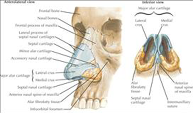
Picture 1 . Structure bone And cartilage nose 6
The nasal septum is a partition that divides the nasal cavity into two nasal cavities and is anatomically formed by bone and cartilage structures. The bone parts are the perpendicular lamina of the ethmoid bone, vomer, and the nasal crest of the maxillary bone. And Christ nasal OS palatine. Whereas part bone vulnerable is septal cartilage, lamina quadrangular And columella. Septum coated by perichondrium in the section bone vulnerable And periosteum in the section bones, while in outside lined by nasal mucosa. 5-7
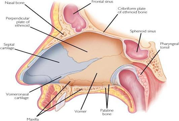
Back rice is part Which most stand out on face And most often experience trauma. Septal abnormalities more easily seen in Caucasian races with the form nose Which more tall compared to with race Asia or Africa. While in children under 5 years old, septal abnormalities are not easily visible because the nose is not the most prominent part of the child's face. 5-7
The maxilla anteriorly and the palatine bone posteriorly border the quadrilateral cartilages. in anteriorly and the vomer posteriorly. The junction between the maxillary and palatine bones forms a prominence, to which the quadrilateral cartilages are attached by fibrous tissue. The junction between the vomer and maxillary bones, in early development, is connected by fibrous tissue, which then becomes bone tissue . The anterior end of the perpendicular plate of ethmoid is a groove where the nasal process of the frontal bone and the nasal bone are attached. The lower end lies in the groove on surface superior from vomer, when join with septum is the thickest place and there are no curves. 5-7
Nose outside receives blood supply from the facial artery which originates from the superior vena cava artery. external carotid and ophthalmic arteries which arise from the internal carotid artery. The inner nose receives blood from the internal carotid artery and the external carotid artery. The part lower nose get bleeding from branch a. maxillary internal among them end a. palatine major And a. sphenopalatine. Part front nose receives blood from the branches of the facial artery. 8,9On part front septum there is anastomosis from branches a. sphenopalatine, a. ethmoidalis front (branch from a. ophthalmic ), a. labial superior and
a. palatine major, Which called plexus Kiesselbach ( little's) area ). Plexus Kiesselbach's nodule is superficial and easily injured by trauma, so it is a frequent source of epistaxis. Veins nose have Name Which The same And walk adjacent to the artery. 3,10
The innervation of the nose comes from branches of nerve fibers. The upper outer surface is innervated by the supratrochlear and infratrochlear nerves, while part inferior get innervation from branch nerve infraorbital And anterior ethmoidal nerve. The inner nose is innervated by the anterior ethmoidal ganglion and the sphenopalatine ganglion. 3,9,10
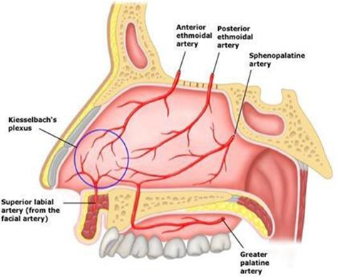
Picture 3 . Circulation blood septum 5
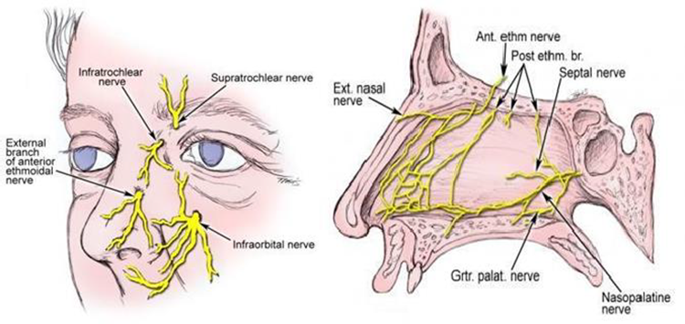
Picture 4. Nerves nose 9
Epidemiology
In general, nasal bone fractures are the most common fractures in the facial area and are the third most common fractures in all fractures in the human body. In the United States, nasal bone fractures are the most common facial fractures, accounting for 39-45% with an estimated 50,000 incidents. fracture OS nasal happen every the year. Number incident on man reported two
up to three times more than women. The incidence of nasal bone fractures increases in age 15-30 year Which due to by fight 34%, accident traffic 28% and sports injuries 23%. In children, the incidence of nasal bone fractures and mandible is fracture Which most Lots found with The most common causes are falls, sports activities, accidents, and violence. 3,8,11
Etiology
Fracture OS nasal on mature can due to by fight, injury due to sports, falls and traffic accidents. While in children it is caused by falls while playing and exercising. It should be remembered that nasal bone fractures in children require consideration of child abuse. 3,8,
Pathophysiology
By understanding the pathophysiology of nasal trauma, it is hoped that the diagnosis and management of nasal trauma therapy can be more optimal. Nasal trauma resulting from a blow varies depends on : (1) age patient Which very influences the flexibility of the tissue in absorbing the energy from the blow, (2) the magnitude of the force of the blow, (3) the direction of the blow which will determine which part of the nose is damaged, and (4) the condition of the object that causes nasal trauma. 15
The nasal bones and cartilage are susceptible to fracture because the nose is prominent and is a central part of the face, making it less strong against external pressure. As with other facial fractures, young patients tend to experience comminuted fractures compared to adult patients who more often experience complex fracture types. 11
The weakest area of the nose is the cartilaginous framework and the junction of the upper lateral cartilages with the bone and septal cartilage at the maxillary crest. This area is the most common site of fracture or dislocation in nasal fractures. The large forces from various directions will cause bone nose crushed Which marked with deformity C shape on septum nasal. Deformity form C usually started in part under the dorsum
nasal And expand to posterior And inferior around laminate perpendicular OS ethmoid and ends in the anterior arch of the septal cartilage approximately 1 cm above the maxillary crest. Part deviation septum consequence fracture OS nasal can include fracture on cartilage septum nasal. Fracture OS nasal lateral is fracture OS nasal Which most frequently encountered and this type of fracture will cause pressure on the ipsilateral nose Which usually covering half bone nose part lower, process maxillary and piriform margins. 3,11,16
Other traumas that are often associated with nasal bone fractures are frontal fractures, nasoorbitoethmoid fractures, orbital wall fractures, frontal sinus fractures and maxillary fractures. 16
Classification
Based on Direction Trauma
Injury Frontal
Injury frontal generally happen Because caught by a number of big power from the front and is divided into three planes, with the level depending on the strength of the trauma and the extent of the injured area. 3,17
Plane I, which is limited to the tip of the nose ( nasal tip ) and does not extend beyond the anatomical line that separates the lower part of the nasal bone from the nasal spine. With the majority of the impact absorbed by the nasal cartilage, the injury usually involves avulsion of the superior lateral cartilage. Dislocation posterior septum And cartilage alarm Also Possible happen, But unlikely .
Plane II, that is covers spine nasal as well as back rice And septum nose. Injury type This result in bone nose become flat (fracture depression) accompanied by with deviation septum, torn mucosa, And fracture spine nasalis. Fracture and dislocation of the nasal septum indicate a severe injury, with collapse of the dorsal surface of the septum. The nasal septum may be involved in about 20% of all traumatic fractures. nose.
Plane III, that is injury Which caused by strength Which big And the impact can be involving fracture orbit or even expand until to structure in
Injury Lateral
in base skull. On injury frontal severe cause " open-book fracture ", where the septum rice becomes collapsed and the nasal bone was stretched out. However, a greater force will cause comminuted fracture on the nasal bone and even part front The maxillary process becomes flat and the nasal dorsum widens.
The most common type of this injury is due to the absence of structural support on both sides of the nasal pyramid after being hit by a large amount of force from the side. This type of injury can be divided into three planes, with the level depending on the strength of the trauma and the extent of the injured area. 3,17
Plane I, which is only an ipsilateral nasal bone fracture, this is an incident that most general, Which usually produce looks existence depression of two-thirds of the surface of the nasal bones.
Plane II, which can be caused by sufficient force where this second type of injury will involve a contralateral nasal bone fracture. and also fractures of the septum rice. In lateral injuries, nasal septal fractures usually extend posteriorly into the ethmoid bone.
Plane III, which is the third type of injury, with greater force will result in fractures of the maxilla and lacrimal bone, can also result in total dislocation of the nasal architecture, or even injury to the lacrimal apparatus.
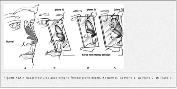
Picture 5. Fracture OS nasal with direction trauma from front. 3
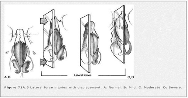
Picture 6. Fracture OS nasal with direction trauma from lateral. 5
Diagnosis
Anamnesis
The time span between the occurrence of trauma and treatment by a head and neck surgeon is the most important factor in patient management. However, it is very important to know the patient's age, the strength of the trauma, the patient's complaints such as epistaxis, nasal congestion, bruising, and a history of trauma or previous nasal surgery. Knowing the time of occurrence trauma as well as estimate direction And the magnitude strength from clash what happens is no less important. For example, trauma from the frontal direction can press the nasal dorsum and cause nasal fractures. Severe edema or hematoma in the soft tissue can obscure the assessment of nasal bone fractures. as well as degrees fracture, so also deformity nose Which happen will makes it difficult to assess old trauma and new trauma so that it will affect the therapy given. 3.16
Complaint clinical like rhinorrhea Which happen in a way Keep going continuously after Nasal trauma should be suspected of causing cerebrospinal fluid leakage caused by a skull base fracture or a fracture of the frontal sinus. 3
Inspection physique
The aim of the physical examination of nasal trauma patients is to establish diagnosis, determine is there any trauma other Which related with Nasal trauma and assessment for case management. Establishing a diagnosis of nasal trauma nasal requires inspection physique Which Good, by Because half from patient trauma Nasal trauma that comes to the emergency room is undiagnosed due to nasal edema often covering trauma to the nasal pyramid area. 13
However, a doctor when treating a patient with a nasal fracture, should not only focus on the traumatized nose, but a thorough examination is very important for patients who have experienced a traffic accident or a fight. A substantial blow to the middle face area will cause trauma to the spine, therefore the doctor must have clinical considerations in taking action without ignoring spinal trauma. Throughout the doctor's initial assessment must ensure that road breath patient safe And ventilation Which optimal. This matter very important to remember the fracture OS nasal often connected with trauma on the head and neck that can affect the airway. 13,18
Inspection side outside And in done For look for existence change shape, shift (deviation) or abnormal shape. The presence of hematoma, laceration and mucosal tears should be suspected of fracture. Eyelid edema, periorbital ecchymosis, ecchymosis sclera, bleeding subconjunctiva And trauma lacrimal are additional clinical signs. Intranasal examination found mucosal decongestion, blood clots, cerebrospinal fluid leakage, deviation or protrusion of the nasal septum. 19,20
On inspection physique with palpation bimanual on nose can crepitation is found, the nasal bone curve is felt and the bone becomes irregular. In patients with septal hematoma, a shiny white or purple area appears that changes on one or both sides of the nasal septum. Delay in identifying And Handling cause deformity nose saddle ( saddle nose ). Anterior rhinoscopy examination must be supported by adequate lighting and cooperative patient conditions. The use of a nasal speculum and headlamp will expand the field of view. 10,16
Fracture OS nasal marked with laceration on nose and also epistaxis due to tearing of the mucous membrane. The soft tissue of the nose will Ecchymosis and edema appear which occur within a short time or several hours after trauma and tend to appear under the nasal bone and then spread to the upper and lower eyelids. Deformity nose like deviation septum or depression back nasal which in new trauma will be very typical, but the deformity that occurred before the trauma often cause mistake on trauma new. Inspection Which thorough on the nasal septum it is very important to determine septal deviation and septal hematoma which are absolute indications for drainage with soon. 16.21
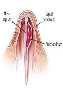
Figure 7. Nasal trauma in children and septal hematoma.
Fracture bone ethmoid usually happen on patient with fracture OS nasal severe fragmentation with the nasal pyramid bone has been pushed back into the ethmoid labyrinth, accompanied by crushing and often with damage to the medial canthal ligament, lacrimal apparatus and cribriform plate causing cerebrospinal rhinorrhea . 10
Inspection support
Supporting examinations in the form of plain nasal photos, Water's and if necessary a CT scan can be performed. However, if there is no suspicion of a nasal bone fracture with complications, inspection radiology seldom recommended Because in reality is less sensitive and specific, so nasal view photos are only indicated if there is doubt in the physical examination performed. Radiography is unable to identify abnormalities in the cartilage and doctors often misinterpret normal sutures as fractures accompanied by dislocation. 10,22,23
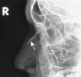
Picture 8 . Photo Lateral Os Nasal. 3
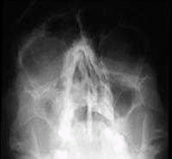
Pictures 9 . Photo Water's 22
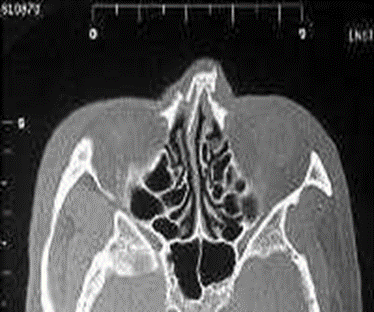
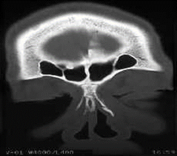
Picture 10 . CT Scan piece axial And coronal head. 22
CT Scan can be used to enforce fracture diagnosis nasal os after it is done inspection previous clinical or if suspected happen other facial trauma. However, when clinical examination finds clinical symptoms such as rhinorrhea cerebrospinal, disorders movement extra eye or malocclusion may indicate a nasal fracture. 3,22-24
Ultrasonography examination is currently being developed as a technology that can be used to diagnose nasal fractures, and several studies have shown that the use of high-resolution ultrasound has better accuracy than CT scans or x-rays and can be used in intraoperative repositioning of nasal fractures. 3
Management
Nasal bone fracture is a facial fracture that is often encountered and if left without proper treatment and within the right time frame, it causes changes in the structure of the nose or soft tissue so that there will be changes in shape and function. 16
Objective Handling fracture OS nasal is overcome condition emergency optimally that is with Handling road breath ( airways ), function breathing ( breathing ) and circulation ( circulation ). Other goals are to restore nasal airway patency, reposition the septum in the midline, maintain the integrity of the nasal cavity, prevent postoperative obstruction, treat septal perforation, prevent changes in the shape of the nasal dorsum, prevent nasal growth disorders and restore a satisfactory appearance. Some treatments for nasal bone fractures are: 3,16,23
Handling in room emergency
The basis for handling trauma patients in the emergency is to address Airway, Breathing, Circulation (ABC) . Patient with Severe nosebleeds are usually controlled by administering topical vasoconstrictors. If the bleeding does not stop, given a ribbon tampon with vaseline, balloon catheterization or ligation of the blood vessel. A ribbon tampon with vaseline is usually placed in the nasal cavity for two days until the bleeding stops. while patient prepared to be done action operation. 16.23
Table 1 . Handling Emergency On Fracture Os Nasal 3 Emergency Management of Nasal Fractures
Condition Emergency | Management |
Bleeding great | Installation tampon, ligation, And cauterization |
Hematoma septum rice | Incision And drainage quick |
Rhinorrhea fluidcerebrospinal | Consulted quick to Neurosurgeon |
Complaint visual | Consulted quick to Ophthalmologist |
Conservative
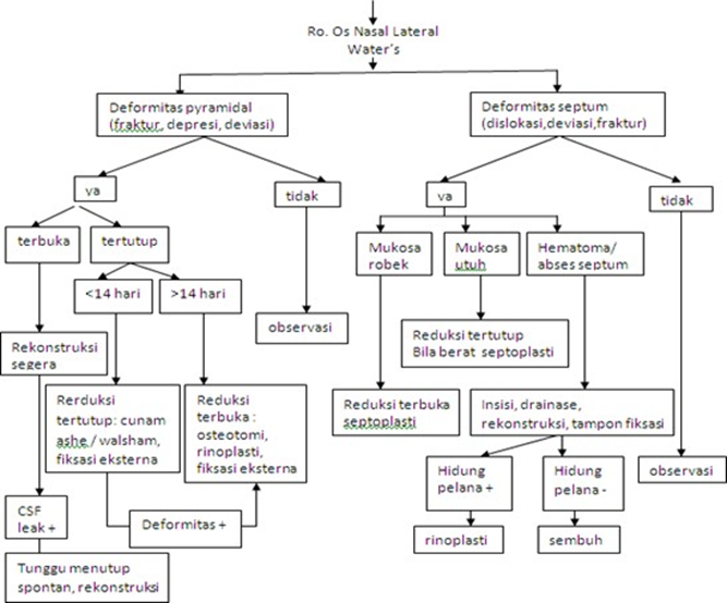
Management of nasal bone fractures is based on clinical symptoms, functional changes And form nose. Lots expert believe that fracture OS nasal type Certain patients are sufficient to receive conservative therapy in the emergency room. In conservative treatment, giving antibiotics aims to reduce the risk of infection and complications. Giving antibiotics therapy analgesics play a role to reduce complaint painful and provide comfort to the patient. 2,3,16
Prognosis
Most undisplaced nasal bone fractures will heal without cosmetic or functional deformity. Open and closed reduction techniques will reduce the cosmetic and functional deformity in patients. Reductions that result in malunion or deformity may require further reconstructive reduction, depending on the severity of the injury and the difficulty in performing the reduction. reduction on injury main. Septorhinoplasty still become standard treatment for less than satisfactory results and is often necessary in cases of failed reduction. 24
REFERENCE
LIST LIBRARY
Donald PD, Sykes J. Facial Fracture. In: Ballenger JJ, Snow JB. Ballenger's Otolaryngology Head and Neck Surgery. 6th ed. Hamilton: BC Decker Inc; 2003. p.900-46.
Michael F Zide. Nasal and Nasoorbital Ethmoid Fractures. In: Dina KR, Delois P, Darlene BC, editors. Principles of Oral and Maxillofacial Surgery. Philadelphia: Lippincott; 1992. p. 547-57.
Grant S, Carlos M. Nasal Fracture. In: Byron J Bailey, et a. Otolaryngology Head and Neck Surgery. Philadelphia; Lippincott Williams & Wilkins: 4th ed . 2006. 71A. p996-1008.
Tardy ME. Surgical Correction of Facial Deformities. In Ballenger JJ. Diseases of the Ear, Nose, Throat, Head And Neck. Translation Staff Expert Part ENT RSCM-FKUI. Jakarta: Binarupa Aksara. 1994. p.28-99.
Walsh WE, Korn RC. Sinonasal Anatomy, Function, And Evaluation. In: Bailey BJ, Johnson JT. Head and Neck Surgery-Otolaryngology. 4th ed, Vol.1. Philadelphia: Lippincott Williams & Wilkins; 2006. p.307-19.
Weller MD, Drake AB. A Reviews of Nasal Trauma. Brit Med J . London. 2006; 8
: 21-8. (accessed date 22 May 2015). Downloaded from http://www.amedeo.co.id
Ballenger JJ. Anatomy and Physiology Of The Nose And Paranasal Sinuses. In: Ballenger JJ, Snow JB. Ballenger's Otorhinolaryngology Head And Neck Surgery.6th ed. Hamilton: BC Decker Inc; 2003. p.547-60.
Ross Adam T, Nasal and Septum Frakturs.2013. (accessed 21 July 2015) Downloaded from http://emedicine. medscape.com/article/878595-overview#showall.
Mountain Roudney E. Surgical Correction of Nasal Fracture. Bleach Nigel et all. In Operative Otorhino-Laryngology. Oxford; Blackwell Science Ltd: 1997. 22.p. 161-164.
Brian P, Kelley BS, How to R, Samuel S. Evaluation and Reduction of Nasal Trauma. Journal of Plastic Surgery, Facial Trauma. 2010; 339-47. (accessed on May 22, 2015). Downloaded from: http://www.medscape.co.id.
Mendez DR, Lapointe A. Nasal Trauma and Fractures in Children. Available at. (accessed July 21, 2015). Retrieved from: http://www.uptodate.com
Anderson PJ. Fractures Of The Facial Skeleton In Children. J Otolaryngol Head and Neck. 1995; 26:47
Maniglia AJ, Kline SN. Maxillofacial Trauma In The Pediatric Age Group. Otolaryngol Clin North Am 1983; 16:717
Perkins SW, Dayan SH, Sklarew EC, et a. The Incidents Of Sports-Related Facial Trauma in Children. Ear Nose Throat J 2000; 79:632.
Gregory Staffel. Nasal Fracture. Current Therapy in Otolaryngology–Head and Neck Surgery. 6th ed. Saint Louis; Mosby Company: 1998. p.133-4
Chegar BE, Tatum SA. Nasal Fracture. In: Cumming CW, et all. Cumming Otolaryngology Head and Neck Surgery. 4 th ed. Baltimore; Mosby Inc: 2005. Chapter 41.
Stranc MF, Robertson GA. A Classification Of Injuries Of The Nasal Skeleton. Ann Plast Surg. 1979; 2.468
Weller MD, Drake AB. A Reviews of Nasal Trauma. Bri Med J. London 2006; 8 : 21-8. (accessed date 22 May 2015). Downloaded from http://www.amedeo.co.id
James KP, Robert MB. Nasal Fractures. In: Raymond JF, Robert VW, editors. Oral and Maxillofacial Trauma. Philadelphia: WB Saunders; 1991. p.600-15
Brian R, Bradley S. Management of Nasal Fractures. Arch Family Med. 2000; 9: 738-42. (accessed May 22, 2015). Downloaded from http://www.amedeo.co.id
Kucik CJ, et al. Management of Acute Nasal Fractures. In AAFP Journals. 2004; 70(7): 1315-20.(accessed date May 2 2015). Downloaded from http://www.BMJ.com
Smith JE, et al. Nasal Fracture Imaging. (Accessed July 19, 2015). Retrieved from http: //emedicine.medscape.com /article/ 391863-overviewl.
Daniel SY, Yea SH, Park HJ. A New Approach to the Treatment of Nasal Bone Fracture. Radiologic Classification of Nasal Bone Fracture and Its Clinical Application. J Oral Maxillofac Surgery. 2011; 69: 2841-47
Lalwani AK. Nasal Trauma. Current Diagnosis and Treatment Otolaryngology Head and Neck Surgery. Second ed. New York; Lange Mc Graw-Hill Company: 2007. Chapter 11.
Soetjipto D, Wardhani RS. Facial Trauma. In the Plastic and Reconstruction Study Group. Guideline for ENT-KL Diseases in Indonesia. Jakarta: 2003. p.82.
Kentjono WA. Management of Maxillofacial Trauma. PKB IX Health Sciences ENT-KL. Emergency Management in the Field of Ear, Nose, Throat-Head and Neck Surgery. Editor Ario Kentjono Widodo et al. Department/SMF Health Sciences ENT-KL FK UNAIR-RSUD Dr. Soetomo Surabaya: 2011. pp.165-181.
Corry JK, Timothy C, James P. Management of Acute Nasal Fractures. Am Fam Physics 2004; 70(7): 1315-20
Green KM, Board T, Mason JT. Alar Haematoma. J Laryngol & Otol 1999; 113: 1104-5. (accessed May 22, 2015). Downloaded from http://www.amedeo.co.id
Murphy J, Marshall AH, Jones NS. Restoration of the Impacted Nasal Pyramid Using Kirschner Wire. J Laringol & Otol 2004; 118: 543-5. (accessed 22 May 2015). Downloaded from http://www.amedeo.co.id
Richard LA, Robert H. Nasal Fracture. Cummings Otolaryngology Head and Neck. Chapter 42. 1990. p.1-17
Wang FG, Kern EB. Nasal Fractures. In: Gates A. Current Therapy In Otolaryngology: Head And Neck Surgery. Philadelphia. PA; BC Decker: 1990: 105-109.
Kim JE, Park PH, Yoon CH. Analysis Of Nasal Septum Fracture Com- Bined In Nasal Bone Fracture Using Cr. J Korean Soc Plast Reconstr Surg 1998;25:852.
Rhee SC, Kim YK, Cha JH, et a. Septum Fracture In Simple Nasal Bone Fracture. Plast Reconstructive Surgery 2004;113:45-52.
Gerd JR, Carsten CB, Milo F, Jorg S. Technique and Timing for Closed Reduction of Isolated Nasal Fracture. A Retrospective Study. Ear Nose And Throat. J New York. 2002; 81(1):49-54
Zargar M, Khaji A, Karbakhsh M, Zarei M. Epidemiology Study Of Facial Injuries During A 13 Month Of Trauma Registry In Tehran. Indian J Med Sci 2004; 58: 109-13
Comments