ABSCESS SUBPERIOSTEAL AS COMPLICATIONS SINUSITIS MAXILLARY DENTOGEN IN GERIATRIC PATIENTS
I Made Nudi Arthana
Sinusitis maxillary dentogen generally happen unilateral, with incident reach 25-75% of all cases of chronic maxillary sinusitis 4,6 . Clinically, the symptoms of dentogenic maxillary sinusitis can resemble the symptoms of other rhinosinusitis. However, late diagnosis and improper treatment can lead to serious complications, namely spread to the orbital or intracranial region 4 .
LITERATURE REVIEWS
Anatomy Nose And Sine Paranasal
The nose and paranasal sinuses are a functional unit that is part of the tract respiratory. Nose consists of from part external And cavity rice. Part The external part of the nose consists of 2 parts, namely the pars osseus and pars cartilaginous. In the Inferiorly there are two orifices called nares. The osseous part consists of the nasal bone, the frontal process of the maxillary bone, and the nasal part of the frontal bone. The cartilaginous part consists of 5 main cartilages, namely 2 pieces cartilage lateral, 2 fruit cartilage style rice, And cartilage septum. Muscle main from The external parts of the nose are the nasalis muscle, the dilator naris muscle, and the levator labii superioris muscle. style rice. Third muscular This innervated by nerve facialist (CN VII). The vascularization of the external part of the nasal cavity comes from branches of the ophthalmic artery, maxillary artery, and facial artery 12,13 .

Picture 1. Part External Nose 12
The nasal cavity is a triangular shaped structure, extending from the nares anteriorly to the aperture nasal posterior (koana) in part posterior, And separated by septum nasal in the middle. Its superior boundary is the lamina cribriformis and its inferior boundary is the palate. hard Which consists of from process palatine OS maxillary And pars horizontal OS palatine. The posterior part of the nasal cavity opens into the nasopharynx. The nasal cavity is lined with mucosa, except in the vestibule which is lined with hairy skin 10,12,13 .
On wall lateral cavity rice there is 3 projection bone, that is concha superior, media, and inferior. Sometimes can found concha fourth Which called concha suprema 10 . Superior concha And concha media is part from OS ethmoid, whereas concha inferior is separate bone 13 . Part front concha media oriented vertical, attached in superior on lateral margin laminate cribriform. Part posterior concha media attached on laminate papyracea and the medial wall of the maxillary sinus. The tip of the inferior concha terminates about 1 cm anterior to the pharyngotympanic tube 10 . In the inferior part of each turbinate there is a gap called the meatus, so that there are inferior, middle and superior meatus 12,13 .
The superior meatus plays a role in the drainage of the posterior ethmoid sinus. Drainage from the sphenoid sinus is through the sphenoethmoidal recess which is located superoposterior to the superior concha. The middle meatus is located inferior to the middle concha, and is the opening of the frontal sinus, maxillary sinus, and anterior ethmoid sinus 10,12 . The inferior meatus is a horizontal slit that located inferolateral from concha inferior. On meatus inferior there is estuary from nasolacrimal duct which drains tears from the lacrimal sac 12 .
In the middle meatus there is a functional unit known as the osteomeatal complex, which consists of the maxillary sinus ostium, ethmoid infundibulum, uncinate process, hiatus semilunaris, middle meatus, and ethmoid bulla 10,11 . The ethmoid infundibulum is a funnel-shaped gap in the anterosuperior part of the middle meatus. The infundibulum is connected to the frontal sinus via the frontonasal duct. Drainage from the frontal sinus empties into a semicircular depression located at the superomedial border infundibulum, Which called hiatus semilunar 11,12 . Hiatus semilunar located between limit front bull ethmoid And process uncinate 10 . Bula ethmoid is the largest anterior ethmoidal cells located superior to the hiatus semilunaris, and are an important anatomical landmark 11,12,13 .
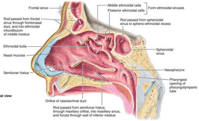
Picture 2. Wall Lateral Rice Cavity 12
The medial part of the nasal cavity is the nasal septum, which has the main components of the perpendicular part of the ethmoid bone, the vomer, and the septal cartilage 12 . The perpendicular part of the ethmoid bone forms the superior part, the vomer forms the posteroinferior part of the septum, and the septal cartilage is found in the anterior part of the septum 13 .
In general, the nasal cavity is innervated by branches of the trigeminal nerve (CN V), namely the ophthalmic nerve (V1) and the maxillary nerve. (V2) 13 . The posteroinferior part is innervated by the nerve nasopalatine And nerve palatine major Which is branch from nerve maxillary. While the anterosuperior part is innervated by the anterior and posterior ethmoidal nerves, branches of the nasociliary nerve which is a branch of the ophthalmic nerve 12. Olfactory nerve (CN I) Which play a role in function smell, originate from cell on epithelium The olfactory bulb is located in the superior part of the nasal cavity, then runs superiorly through the cribriform lamina towards the olfactory bulb 12,13 .
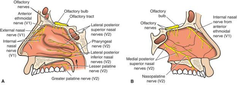
Picture 3. Nasal Cavity Innervation 13
The main vascularization of the nasal cavity comes from the branches of the maxillary artery which is a branch of the external carotid artery, and the ophthalmic artery which is a branch of the internal carotid artery. The nasal septum is vascularized by anastomoses from several blood vessels, namely the anterior ethmoidal artery, posterior ethmoidal artery, sphenopalatine artery, greater palatine artery, And labial artery superior. Anastomosis vessels This blood is known as the Kiesselbach plexus 12 .
Flow vein from part external nose part big flow to vein facialist through the angular and lateral nasal veins. Venous drainage from the nose is through the sphenopalatine, facial, And ophthalmic, going to A plexus vein submucosa. Plexus This is an important part of the body's thermoregulatory system, exchanging heat and warming the air before it enters lungs. Plexus This relate with sine cavernosus so that located in "area danger" on face. Drainage lymphatic cavity rice going to gland lymph cervicalis profundus, and in the vestibule 13
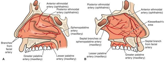
Picture 4. Vascularization Cavity Rice 13
Paranasal sinuses are cavities in the maxillofacial region and skull bones that are filled with air, lined by mucosa, and is connected to the nasal cavity. At In humans, the paranasal sinuses consist of the maxillary sinus, ethmoid sinus, frontal sinus, and sphenoid sinus 11. The ethmoid sinus is formed at birth, while the other paranasal sinuses develop through process pneumatization along with process grow flower. Pneumatization on frontal sinus generally start happen on age 4-8 year, sine maxillary on age 1-4 year, and sphenoid sinus at the age of 1-3 years 10 .
The frontal sinuses are located in the frontal bone, normally there are a pair in adults, but they are not developed in 5% of the population 10 . The frontal sinuses are slightly triangular in shape and are separated by a bony septum in the middle. Drainage from the frontal sinuses is through the frontonasal duct to the ethmoid infundibulum. The frontal sinuses are innervated by branches of the supraorbital nerve (CN V1) 12,13 .
The ethmoid sinuses consist of the anterior, middle, and posterior ethmoid sinuses. The ethmoid sinuses are formed from invagination membrane mucosa meatus media And superior to in bone ethmoid between the nasal cavity and the orbital cavity 12. The lateral boundary of the ethmoid sinus is the lamina papyracea, whereas limit medial that is concha superior. Sine ethmoidalis front located anterior to the basal lamella, while the posterior ethmoid sinus is located between the basal lamella and the sphenoid sinus 10 . Drainage from the anterior ethmoid sinus is towards the middle meatus, while the posterior ethmoid sinus is drained into the superior meatus. Innervation of the ethmoid sinus is by branches of the nasociliary nerve (CN V1) 12 . Some anatomical variations of the ethmoid cells include Haller cells and Onodi cells. The anterior ethmoid cells that are parallel to the floor of the orbital cavity and located lateral to the lamina papyracea are called Haller, whereas cellulae ethmoidalis posterior Which develop And invade The superolateral part of the sphenoid sinus is called Onodi's cell (sphenoethmoidal cell) 10 .
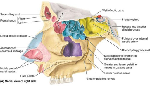
Picture 5. Paranasal Sinuses 12
The sphenoid sinus is located in the corpus of the sphenoid bone, generally bilateral but with asymmetrical size. The sphenoid sinus has extensive pneumatization so that the walls of the sine sphenoid shaped thin And fragile. Sine sphenoid bordering with structure important, including the optic nerve, optic chiasm, pituitary gland, internal carotid artery, and cavernous sinus. Sine sphenoid vascularized by artery ethmoidalis posterior, And innervated by the posterior ethmoidal nerve 10,12 .
The maxillary sinus or Highmore's antrum is the largest paranasal sinus, located in OS maxillary. Sine maxillary shaped like pyramid with apex Which leading to the zygoma and the base adjacent to the nasal cavity. The roof of the maxillary sinus is formed by base cavity orbit, whereas part inferior bordering with process alveolar and the roots of the premolar and molar teeth 10,11 . The anterior wall of the maxillary sinus is formed by the facial part of the maxillary bone, and on its inner edge there is a sulcus through which the anterior superior alveolar artery, vein, and nerve pass. The infratemporal part of the maxillary bone forms the posterior wall of the maxillary sinus, and is the anterior wall of the pterygopalatine fossa 14 .
Fossa pterygopalatine in a way clinical very important Because can become track spread of tumors or infection to the surrounding tissue. The pterygopalatine fossa is a funnel-shaped structure, bounded by the base of the skull superiorly, the posterior part of the oral cavity inferiorly, and the pterygoid processes and the anterior part of the greater ala of the sphenoid bone posteriorly. The pterygopalatine fossa is connected to the orbital cavity through the inferior orbital fissure and to the middle cranial fossa through the foramen rotundum 11 .
The arterial supply to the maxillary sinus comes from branches of the maxillary artery, namely the superior alveolar artery, the infraorbital artery, and the posterior lateral alveolar artery, as well as the greater palatine artery and the descending palatine artery 12,14 . The maxillary sinus is innervated by the superior alveolar nerve which is a branch of the maxillary nerve 12 .
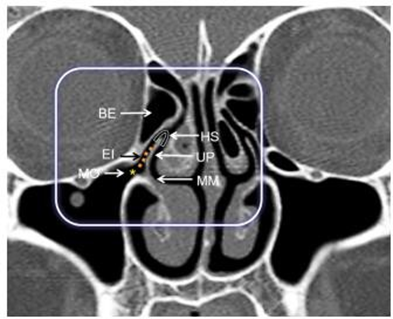
Figure 6. Components of the Osteomeatal Complex on Coronal CT. MO: maxillary sinus ostium; EI: infundibulum ethmoid; BE: bull ethmoidalis; MM: meatus medium; UP: uncinate process; HS: hiatus semilunaris 10
Physiology Nose And Sine Paranasal
The nose plays a role in the function of smell, respiration, and as a pathway for the release of secretions from the paranasal sinuses and nasolacrimal ducts 12 . When air passes through the nose, three respiratory functions normal that different things are done by the nasal cavity: (1) the air is warmed by mucosa concha And septum, Which own total wide surface around 160 cm2 ; (2) humidified air; and (3) air is filtered so that particles larger than 6 micrometers cannot enter the respiratory tract 15 . The paranasal sinuses, in addition to playing a role in respiratory function, also function to reduce the weight of the skull bones and in voice resonance 13 .
Combination from its vastness surface mucosa on cavity rice And concha, secretion mucus, and the abundant blood supply in the lamina propria allows the nose to warm And humidify air Which inspired. Nose can change dry and cold air becomes moist and warm air in less than a second, and the process arrangement temperature air This very important For function And health channel lower respiratory tract (larynx, trachea, bronchi, bronchioles, and alveoli) 11 .
Nose And sine paranasal coated by epithelium layered pseudo ciliated Which have a lot cell goblet, supported by laminate property vascular Which contain gland serosa and mucosa and numerous thin-walled venules. Together, the epithelium and lamina propria form the mucosa. The mucosa lining the nose and paranasal sinuses is attached to the underlying periosteum, and this mucoperiosteum is commonly referred to as the Schneiderian membrane 11 .
Mucociliary clearance is the primary defense mechanism of the respiratory tract to protect against inhaled pollutants, allergens, and pathogens. Its functional components include cilia And hammer mucus Which secreted by cell goblet in epithelium And gland mucus in the laminate property. Mucus act like paper glue, catch particle air Which inspired through the nose. Mucus consists of two layers; a thin, watery sol layer that moistens the cilia so that make it easier its movement, And layer gel Which thick And sticky which plays a role in capturing particles. Cilia act in a coordinated manner to move layer gel And particle Which trapped (with speed around 6 mm per minute) towards the sinus ostium and nasal cavity, then towards the nasopharynx 11 .
The natural ostium of the maxillary sinus is located in the superior part of the medial wall of the maxillary sinus, inferior to the floor of the orbital cavity 6 . The mucus produced by the maxillary sinus mucosa is pushed superiorly by the cilia towards the ostium against gravity 15 .
The hairs in the nasal vestibule are important for filtering large particles. In addition, particle removal also occurs through the turbulent precipitation mechanism. This is when air passing through the nasal passages hits many obstacles, including the conchae, septum, and pharyngeal wall. Each time air hits one of these obstacles, it must change direction its movement. Particles Which floating in air, own mass and momentum Far more big from air, No can change direction journey they as fast as air. Therefore, the particles are then trapped in the mucus layer and pushed by the cilia into the pharynx 15 .
Sinusitis Maxillary Dentogen
Definition
Dentogenic maxillary sinusitis or odontogenic sinusitis is bacterial maxillary sinusitis, with or without distribution to sine paranasal other, Which happen consequence infection adjacent teeth or complications from the procedure on teeth 2,3 .
Epidemiology
Approximately 30 million people in the United States suffer from rhinosinusitis 17 . Maxillary sinusitis dentogenetic generally happen unilateral (92.6%), with incident reach 25- 75%
from all over case sinusitis maxillary chronic. Only 7.4% case sinusitis maxillary dentogen occurs bilaterally. In the literature it is stated that 45-75% of maxillary sinusitis unilateral is dentogen maxillary sinusitis 4,6,19 . Dentogen maxillary sinusitis occurs most often in the 40-60 year age group, with a prevalence of women (57.7%) higher than men (42.82%) 2 .
Etiopathogenesis
The most common etiology of dentogenic maxillary sinusitis is dentoalveolar procedures or odontogenic infections with perforation of the Schneiderian membrane. Dentoalveolar procedures such as tooth extraction, dental implant placement, endodontic procedures, and others can cause perforation of the Schneiderian membrane or oroantral fistula formation that predisposes to infection in the maxillary sinus. Odontogenic infections such as periodontitis, periapical infections, or infections of the tooth roots are also at risk of causing dentogenic maxillary sinusitis 5,27 .
Pathogenesis from sinusitis maxillary dentogenetic Also influenced by proximity anatomically between the tooth root and the floor of the maxillary sinus 2 . Anatomically, the floor of the maxillary sinus is connected to the alveolar process in the maxilla. The average distance between the root of the molar tooth and premolar that is 1.97 mm, However distance This varies greatly from person to person individual 27 . Root molar teeth second (M2) generally most near with base sine maxillary, followed with first molar (M1), third molar (M3), second premolar, and first premolar 4.6 .
In addition, under the maxillary sinus mucosa there are branches of blood vessels, nerves, and lymph vessels that supply the tooth roots. So that the infection can spread to the maxillary sinus through the blood circulation because of the similarity of vascularization between the sinus mucosa and periodontal tissue, and/or through the facial space 2,6 .
Process inflammation on sinusitis maxillary dentogenetic can differentiated become two different stages: the acute or invasive stage characterized by the activation of non-specific immunity, mainly characterized by neutrophils and macrophages, and followed by the chronic stage, which shows the characteristics of adaptive immune reactions. During the acute phase, bacteria can spread to network Which close together in a way direct, stimulate epithelium membrane And trigger hypertrophic reaction 2.6 .
Odontogenic infections begin with bacteria attaching to the outer surface of the tooth, damaging the outer enamel and inner dentin and penetrating the pulp. Once the infection enters the pulp, necrosis and pus formation occur. The body is unable to eliminate the source of infection. Because pulp necrotic protected in in root tooth. Bacteria to colonize part root apical and bacterial toxins can damage tissue, causing periapical infection 5 . If the infection process spreads to the Schneiderian membrane, it can cause inflammation and hypertrophy, resulting in the release of pro-inflammatory factors that cause edema, fibrosis, and cystic degeneration. In addition, bacteria from the pathological process of the tooth, through microbial toxins, can strengthen inflammatory mediators such as TNF-α, interleukin (IL)-8, and IL-13, potentially causing changes in ciliary activity 2 . In other studies, the dominance of lymphocytes and plasma cells was found in the inflammatory process of dentogenic maxillary sinusitis, with the activity of inflammatory mediators such as IL-17, IL-6, IL-10, IL-27, and increased levels of IFN-ɣ 6 .
The microbiome in dentogenic maxillary sinusitis is different from other rhinosinusitis. Infection generally is infection polymicrobial, that is existence involvement bacteria aerobic and anaerobes from the oral cavity and from the upper respiratory tract, with a predominance of anaerobic bacteria 5,6,16 . Aerobic bacteria that can be found in cases of dentogenic maxillary sinusitis include Streptococcus alpha hemolyticus , microaerophilic streptococcus, Staphylococcus aureus , And Streptococcus pyogenes . Whereas bacteria anaerob Which general obtained among others Peptostreptococcus , Fusobacterium sporulatum , Propionibacterium acne, And Prevotella spp. More than 158 species of bacteria and several species of fungi such as Candida spp. have been identified as causes of dentogenic maxillary sinusitis 2,16 .
Recent hypotheses are being widely studied regarding the role of bacterial biofilms in the pathogenesis of dentogenic maxillary sinusitis. Bacterial biofilms are dynamic polymicrobial communities of multiple replicating bacteria that have constant metabolism and are incorporated into a matrix rich in exopolysaccharides, proteins, and nucleic acids. Bacterial biofilms are involved in many chronic infections and are more difficult to eradicate due to their structure. Which layered. Layers on biofilm protect microbes in inside from action antibiotics, detergent, or agent antimicrobial other, so that cause infection chronic recalcitrant 2 .
Symptom Clinical and Diagnosis
Symptoms of maxillary sinusitis can be divided into sinonasal symptoms and symptoms of tooth Symptoms sinonasal main Which related with sinusitis maxillary dentogenetic is painful or pressure on the face, nasal congestion, purulent rhinorrhea which is usually unilateral, cacosmia or the perception of smelling a foul odor, bad taste in the mouth and postnasal drip . Based on the literature, the most common symptoms complained of by patients are purulent rhinorrhea, followed by with painful cheek And kakosmia 5.27 . Will but, symptoms This No pathognomonic for dentogen maxillary sinusitis only. Some patients do not complain of nasal odor or may have experienced hyposmia so they do not complain of kakosmia 3 . Some patients with dentogen maxillary sinusitis do not experience any symptoms (asymptomatic) 2 .
Dental symptoms include a history of toothache, loose teeth, gum abscess on the side affected by maxillary sinusitis, or a history of dentoalveolar procedures 6,17 .
On physical examination, anterior rhinoscopy can find purulent secretions in the nares, which are generally greenish yellow and have a foul odor. Pain can be found during palpation of the anterior wall of the maxillary sinus. Nasoendoscopy can show purulent secretions in the middle meatus accompanied by hyperemia and congestion of the surrounding mucosa. Purulent secretions can also be found flowing on the posterior pharyngeal wall 2 . Polyps can also be found in the nasal cavity during nasoendoscopy 3 . On clinical examination of the mouth, cavities, residual tooth roots, pain during tooth percussion, or redness and swelling of the gums can be found 6,16 .
Radiographic examination is an important examination for the diagnosis and management of sinusitis. maxillary dentogen. Radiography periapical And panoramic useful For determine the size of the periapical lesion, visualize pseudocysts, displaced roots, and other teeth or foreign bodies in the in sine maxillary. However, second test diagnostic This is imaging two-dimensional and therefore not specific enough to clearly evaluate the maxillary area compared to three-dimensional techniques, such as computed tomography (CT) 3 .
CT scan is considered the gold standard for adequate imaging of the maxillary sinuses because of its high resolution, ability to differentiate bone and soft tissue, and ability to detect sinonasal inflammation 3 . CT scan examination in dentogenic maxillary sinusitis can show description thickening mucosa, air-fluid level , or unilateral obscuration. Mucosal thickening of more than 2 mm is considered significant 6,16

Picture 7. CT Scan on Sinusitis Maxillary Dentogen with Image of Occlusion of the Left Maxillary Sinus Accompanied by Periapical Abscess in Left 1st Molar Tooth 5
In diagnosing odontogenic maxillary sinusitis, in addition to the pathological picture in the maxillary sinus, it is necessary to identify odontogenic lesions on radiological examination 6 . Several criteria have been proposed by experts regarding the diagnosis of dentogenic maxillary sinusitis. One of them is the endo-antral syndrome criteria, namely 2 :
Pathology pulp on tooth;
Description radiolucent on area periapical on tooth with condition pulp pathological;
The disappearance laminate Dura on base wall sine maxillary Which related to pathological teeth;
Description radiopaque supraapical Which invade sinus cavity maxillary;
Description radiopaque with degrees Which varies in the cavity maxillary sinus 2 .
Lin et al. proposed several criteria to help establish the diagnosis of dentogenic maxillary sinusitis. These criteria are: 6
Patient with clinical symptoms of sinusitis maxillary, with or asymptomatic oral.
There are pathological teeth in the upper row of teeth the same side as the occurrence of maxillary sinusitis, with lesi periapical or the disappearance bone alveolar Which critical (absorption up to 2/3 of the root or more) on cone beam computed tomography (CBCT).
Foreign body in the maxillary sinus or ipsilateral oroantral fistula on the same side maxillary sinusitis
Description CT/CBCT show air-fluid level or opacification sine maxilla or thickening mucosa sine maxillary (> 2 mm) limited on lesi oral ipsilateral in same side as maxillary sinusitis, with features:
Discontinuity base sine maxillary between both of them.
Layer thin basic bone sine maxillary remaining in between the two.
Base sine maxillary Which thick Which thick between both of them.
Definitive evidence of dentogenous maxillary sinusitis is if the patient meets criteria 1, 3, or 1, 2, 4a. Potential dentogenous maxillary sinusitis is if the patient meets criteria 1, 2, 4b. Whereas sinusitis maxillary dentogenetic considered doubtful if it meets criteria 1, 2, 4c 6 .
Management
Once the diagnosis is established, management of odontogenic maxillary sinusitis requires collaboration between specialist doctors. ENT-BKL with a dentist. Successful management of dentogenic maxillary sinusitis involves a combination of definitive therapy of the pathological condition tooth, therapy medication, And therapy surgery 5.17 . Objective from governance sinusitis maxillary dentogenetic that is eliminate reason, reduce symptom And restore normal function of the sinuses 18 .
Medication
Non-surgical management is the first and most important step in the management of dentogenic maxillary sinusitis. The recommended therapy is the administration of antibiotics, intranasal corticosteroids, decongestants and nasal irrigation, which help reduce patient symptoms. Decongestants are given to reduce nasal congestion and open the sinus ostia, but must be given with caution because they can cause a rebound effect . Intranasal corticosteroids are recommended to reduce mucosal edema. Nasal irrigation is recommended 3 times a day for 2-3 weeks 6,18 .
Antibiotics should be given according to culture results, but if not available, ampicillin or piperacillin combined with a β-lactamase inhibitor is recommended 2,4 . Metronidazole may also be considered for eradication of anaerobic bacteria 6,7 . Antibiotics are given intravenously if there is a risk of intracranial or orbital spread of infection 18 .
Choice therapy empirical other is combination levofloxacin And vancomycin. Alternatives include tetracycline, fluoroquinolone, or moxifloxacin. Moxifloxacin has been shown to be an effective agent. antibacterial Which Enough active to anaerob And own activity wide to aerobic gram positive And grams negative. Duration giving antibiotics in sinusitis maxillary dentogen at least 14 day or at least 7 day after symptom missing 2 . Not yet there is consensus what is certain regarding the duration of conservative therapy, with some variations in the literature, namely 2-6 weeks 6,18 .
Administration of systemic corticosteroids, either oral or intravenous, can be useful for reducing inflammation and edema, but must be given with caution because it can cause immunosuppression 7 . Administration of systemic corticosteroids after surgery can reduce edema mucosa, adhesion, closed ostium sine paranasal, as well as reduce postoperative pain. Systemic corticosteroids also shorten the duration of hospitalization 23 .
Surgery
Surgery indicated If symptom settle down after therapy conservative during 48 hours, there is a picture of obstruction in the osteomeatal complex, and mucosal thickening reaches more than half of the maxillary sinus 5,19 . Antibiotic therapy alone is often ineffective in most cases of dentogenic maxillary sinusitis 4 .
There is a number of approach surgery For governance sinusitis maxillary dentogen like surgery sine endoscopic functional (BSEF) And operation Caldwell Luc 27 . Technique Caldwell-Luc is Wrong One technique Which Lots chosen in management of maxillary sinus disease 4. This technique has the advantage of wider visualization, which allow For extraction object foreign, cyst, And tumor on sine maxillary. However, there are some post-operative complications such as post-operative facial swelling, paresthesia of the face or teeth, and sclerosis maxillary sinus wall 6.27 . BSEF is currently widely recommended in management of sinonasal diseases, including sinusitis maxillary dentogen 27 . Based on level infection odontogenic, procedure BSEF can extend from the unilateral maxillary sinus to the ethmoid sinus, even the sphenoid sinus and frontal sinus, with use visualization endoscope 0 degrees. With middle meatal antrostomy , ostium sine maxilla can enlarged so that expected can return
drainage and sinus ventilation 6 .
Indications for BSEF are failure of conservative therapy, chronic odontogenic sinusitis, complications of rhinosinusitis (intraorbital and intracranial complications), benign paranasal sinus tumors, dacryocystostomia, transnasal hypophysectomy, orbital tumors, trauma cases, and management of epistaxis 18 .
Compared with the Caldwell-Luc technique, endoscopic sinus surgery has obvious advantages, such as minimal incision, no scars in the oral cavity, shorter hospital stay, preservation of maxillary sinus functionality, and a much lower complication rate 2 . However, there are some important anatomical structures that at risk experience injury consequence action This, like region orbit, nerve optician, base cranii, internal carotid artery, anterior ethmoidal artery, and others 6 .
Complications BSEF shared become complications major And complications minor. Minor complications including synechiae, bleeding or damage channel water eye, Which can causing epiphora or permanent blockage of the tear duct, double vision due to injury to the eye muscles rectus medialis, emphysema orbital consequence damage on laminate papyracea, And loss of sense of smell. Major complications include severe injury to the anterior and posterior ethmoid arteries, artery carotid internal (Which can cause the bleeding that threaten soul), nerve optician, region orbit And Contents cavity orbit (hematoma retrobulbar, emphysema periorbital), and dura. Cerebrospinal fluid leaks and other intracranial complications may also occur 18 .
Collaboration with Doctor Tooth
Handling to condition pathological in tooth play a role very important in sinusitis management maxillary dentogen 4 . Therapy definitive in field tooth depends on condition underlying pathology, including root canal treatment , periodontal treatment, extraction of teeth that are focal infections, and closure of oroantral fistulas. Dental hygiene and education about dental and oral hygiene are essential 5,18 .
Complications And Prognosis
Dentogenic maxillary sinusitis can cause spread of infection to extrasinus regions. that is orbit, intracranial, And bones. On study Craig et et al., complications orbital complications are the most common (69%), followed by intracranial complications (19%), orbital and intracranial (8%), as well as bone (4%). Complications orbital covering cellulitis or abscess preseptal, cellulitis postseptal (orbital), abscess subperiosteal, abscess orbital (intraconal), And thrombosis cavernous sinus. Complications intracranial including meningitis, cerebral, abscess (epidural, subdural, or intraparenchymal), or cerebral venous sinus thrombosis. Bone complications include osteomyelitis of the sinus wall which can lead to subcutaneous subperiosteal abscess, sinocutaneous fistula, or orbital or intracranial spread 19 .
The prognosis of dentogenic maxillary sinusitis is influenced by the underlying etiology. Generally, a combination of rhinologic and dental therapy provides a good prognosis for patients 6 . A multidisciplinary approach tailored to each patient's condition is essential for successful resolution of the disease 17 .
Complications Orbital Consequence Rhinosinusitis
Spread of infection from the paranasal sinuses can cause serious orbital complications 9 . Orbital complications of rhinosinusitis generally originate from the ethmoidal or maxillary sinuses, less commonly from the frontal sinuses 21 . The main etiopathogenesis of infection in the periorbital area is the spread of infection from surrounding structures, especially the paranasal sinuses 8 . In general anatomically, the lamina papyracea thin, closeness anatomy between orbits And sinuses, and the absence of venous valves in the orbital and paranasal sinus vessels facilitates the retrograde spread of infection. A narrow osteomeatal complex, diabetes mellitus, and immunocompromised conditions also increase the risk of infection 22 .
Orbital complications of rhinosinusitis can be classified into 2 main categories, namely preseptal (periorbital) And postseptal (orbital) 7 . Classification This Then regrouped based on degrees distribution infection. Classification Chandler is system classification Which most general used, Which share complications orbital from rhinosinusitis become 5 groups, namely preseptal (periorbital) cellulitis, orbital cellulitis, subperiosteal abscess, orbital abscess, and cavernous sinus thrombosis 20 .
Cellulitis preseptal (periorbital)
On cellulitis periorbital, infection limited on network soft front from septum orbital and does not pass through the orbital septum. This condition is also known as preseptal cellulitis. Patients typically present with erythema, periorbital edema, and pain. Eye movements are usually intact, and visual acuity is not affected 8,23 .
Cellulitis orbit
Orbital cellulitis involves inflammation of the orbital tissues posterior to the orbital septum. Orbital cellulitis causes diffuse inflammation of the orbital connective tissues. Clinical manifestations of orbital cellulitis include edema with ill-defined borders of the orbital muscle. extraocular, proptosis, chemosis conjunctiva, restrictions movement eye, And painful with or without decline aquity visual. On condition This Not yet obtained formation abscess 8.9 .
Abscess subperiosteal
On abscess subperiosteal, happen formation pussy between wall periorbital And orbital bone 22. Clinically the symptoms are unilateral swelling of the upper eyelid and/or lower, erythema, painful And movement eye Which limited, diplopia, exophthalmos, eyelid only Can opened in a way passive, And chemosis Which significant. Patient suspected experience a subperiosteal abscess if there is worsening proptosis and limited eye movement 25 .
Abscess orbit
Abscess orbit marked with existence pussy in in region orbita. Symptoms clinical abscess orbital, namely unilateral swelling that feels stiff on the upper and/or lower eyelid, erythema, ophthalmoplegia, progressive decrease in vision, and diplopia 8,22,25 .
Thrombosis sine cavernous
In cavernous sinus thrombosis, there is a posterior spread of infection which causes phlebitis on sine cavernosus. In general clinical marked with proptosis, ophthalmoplegia, decline vision, painful head great, fever, change status mental retardation, cranial nerve palsy, and signs of sepsis 22 .
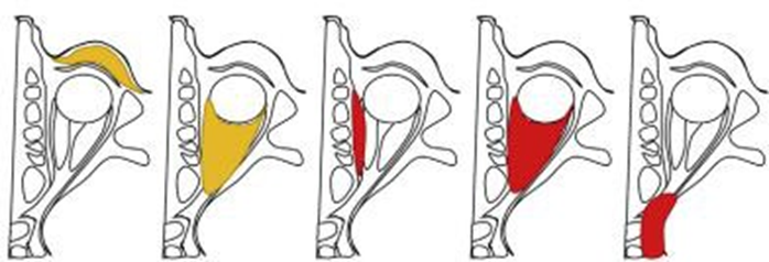
Picture 8. Classification Complications Orbital Consequence Rhinosinusitis Based on Chandler Classification 22
Complications orbital from rhinosinusitis relate with morbidity tall And potential mortality. Therefore, rapid and accurate diagnosis of this complication is essential to provide better outcomes 22 . CT scan is considered the gold standard in diagnostics And For determine wide complications orbital. Inspection This Also allows detection of abscesses 21 .
Governance main from cellulitis preseptal And cellulitis orbital namely giving broad-spectrum oral or intravenous antibiotics, and corticosteroids. Corticosteroids have been shown to be effective in the management of cervicofacial and systemic infections, reducing edema and inflammation in the periorbital and intraorbital regions, and preventing the occurrence of orbital compartment syndrome. Corticosteroid administration has been shown to not only reduce mortality rates, but also reduce the incidence of prolonged neurologic sequelae. Other management includes symptomatic therapy for sinusitis conditions, such as decongestants 21,23 .
In cases of cavernous sinus abscess and thrombosis, in addition to antibiotics, surgical intervention is also required 21 . Some indications for surgery are visual disturbances, signs of orbital abscess, or in patients who do not improve after intravenous antibiotics for 48 hours 22 . The purpose of surgical therapy is to drain pus, open and widen natural ostium of the sinus, and obtain samples for culture examination. Surgery can be performed by an external approach such as an incision on region periorbital, And with approach endoscopic through surgery sine functional endoscopic. Approach Which chosen very depends on condition clinical And consideration of expert surgery. Procedure endoscopic For case abscess subperiosteal generally involving combination of anterior, posterior and ethmoidotomy maxillary antrostomy. In cases where ophthalmoplegia or loss of vision is present, immediate orbital decompression and drainage through an external approach is recommended 25 . If there is a risk of cavernous sinus thrombosis, subcutaneous heparin administration may be considered 21 .
CONCLUSION
Dentogenic maxillary sinusitis or odontogenic sinusitis is bacterial maxillary sinusitis, with or without distribution to sine paranasal other, Which happen consequence infection adjacent teeth or complications from dental procedures. If not treated properly, dentogenic maxillary sinusitis can cause complications in the orbital region due to anatomical proximity. between sine paranasal and orbit as well as absence valves in blood vessels ophthalmic venous blood that allows retrograde spread of infection. Advanced age, uncontrolled diabetes mellitus, and immunocompromised conditions can be predisposing factors for complications. Subperiosteal abscess due to complications of maxillary sinusitis dentogenetic need Handling medication Which combined with surgery. In determining the surgical approach and technique performed, it is necessary to consider the patient's clinical condition to avoid postoperative sequelae. A multidisciplinary approach in collaboration with dentists and other related specialists is essential to provide optimal results and prevent recurrence.
REFERENCE
Fokkens WJ, Lund VJ, Hopkins C, Hellings PW, Kern R, Reitsma S, et al. European Position Paper on Rhinosinusitis and Nasal Polyps 2020. Rhinology. 2020;58(Suppl S29):1-464.
Martu C, Martu MA, Maftei GA, Diaconu-Popa DA, Radulescu L. Odontogenic Sinusitis: From Diagnosis to Treatment Possibilities-A Narrative Review of Recent Data. Diagnostics (Basel). 2022;12(7):1600.
Craig JR, Poetker DM, Aksoy U, Allevi F, Biglioli F, Cha BY, et al. Diagnosis of odontogenic sinusitis: An international multidisciplinary consensus statement. Int Rhinol Allergy Forum. 2021;11(8):1235-1248.
Sabatino L, Pierri M, Iafrati F, In John S, Moffa A, With Benedetto L, et a. Odontogenic Sinusitis from Classical Complications and Its Treatment: Our Experience. Antibiotics (Basel). 2023;12(2):390.
Psillas G, Papaioannou D, Petsali S, Dimas GG, Constantinidis J. Odontogenic maxillary sinusitis: A comprehensive review. J Dent Sci. 2021;16(1):474-481.
Craig JR, Tatar RW, Aghaloo TL, The Truth AT, Gray ST, Mattos JL, et a. Management of odontogenic sinusitis: multidisciplinary consensus statement. Int Forum Allergy Rhinol. 2020;10(7):901-912.
Geusens J, Dubron K, Meeus J, Spaey Y, Politis C. Subperiosteal orbital abscess from odontogenic origin: A case reports. Int J Surg Case Rep. 2020;73:263-267.
Gupta S, Sharma S. Orbital Cellulitis: Multidisciplinary Defining Approach as the Need of the Hour. Indian J Otolaryngol Head Neck Surg. 2019;71(4):464-469.
Sansa Perna A, Gras-Cabrerizo JR, Montserrat-Gili JR, Rodríguez-Álvarez F, Massegur-Solench H, Casasayas-Plass M. Our experience in the management of orbital complications in acute rhinosinusitis. Acta Otorrinolaryngol Esp (Engl Ed). 2020;71(5):296-302.
Vaid S, Vaid N. Sinonasal Anatomy. Neuroimaging Clin N Am. 2022;32:713–734
Whyte A, Boeddinghaus R. The maxillary sinus: physiology, development and imaging anatomy. Dentomaxillofac Radiol. 2019;48(8):20190205.
Moore KL, Dalley AF, Agur AMR. Moore Clinically Oriented Anatomy. 8th Edition. Philadelphia: Wolters Kluwer: 2018.
Winery LE. Snell's Clinical Anatomy by Regions. 10th Edition. Philadelphia: Wolters Kluwer: 2019.
Iwanaga J, Wilson C, Lachkar S, Tomaszewski KA, Walocha JA, Tubbs RS. Clinical anatomy of the maxillary sinus: application to sinus floor augmentation. Anat Cell Biol. 2019;52(1):17-24.
Hall JE, Hall ME. Guyton and Hall Textbook of Medical Physiology. 14th Edition. Philadelphia: Elsevier: 2021.
Micaela CM, Paula CB, David PA. Odontogenic sinusitis, a differential diagnosis: Literature review. World J. Adv Res Rev. 2023;18:1241-1247.
Little RE, Long CM, Loehrl TA, Poetker DM. Odontogenic sinusitis: A review of the current literature. Laryngoscope Investig Otolaryngol. 2018;3(2):110-114.
Rangics A, Répássy GD, Gyulai-Gaál S, Dobó-Nagy C, Tamás L, Simonffy L. Management of Odontogenic Sinusitis: Results with Single Step FESS and Dentoalveolar Surgery. J Pers Med. 2023;13(9):1291.
Craig JR, Cheema AJ, Dunn RT, Vemuri S, Peterson EL. Extrasinus Complications From Odontogenic Sinusitis: A Systematic Review. Otolaryngol Head Neck Surg. 2022;166(4):623-632.
Kim BY, Lee S, Jeon J, Lim IG, Choi GC. Characteristics of Atypical Orbital Complications of Sinusitis. J Craniofac Surg. 2020;31(5):e435-e439.
Welkoborsky HJ, Pitz S, Grass S, Breuer B, Paehler Vor der Holte A, Bertram O, et al. Sinogenic Orbital Complications. Dtsch Arztebl Int. 2022;119:31-37.
Anselmo-Lima WT, Soares MR, Fonseca JP, Garcia DM, Velasco E Cruz A A, Tamashiro E, et al. Revisiting the orbital complications of acute rhinosinusitis. Braz J Otorhinolaryngol. 2023;89(5):101316.
Mahalingam S, Luke L, Pundir J, Pundir V. The role of adjuvant systemic steroids in the management of periorbital cellulitis secondary to sinusitis: a systematic review and meta-analysis. Eur Arch Otorhinolaryngol. 2021;278(7):2193-2201.
Jyani R, Ranade D, Joshi P. Spectrum of Orbital Cellulitis on Magnetic Resonance Imaging. Cureus. 2020;12(8):e9663.
Presutti L, Lucidi D, Spagnolo F, Molinari G, Piccinini S, Alicandri-Ciufelli M. Surgical multidisciplinary approach of orbital complications of sinonasal inflammatory disorders. Acta Otorhinolaryngol Ital. 2021;41(Suppl. 1):S108-S115.
Zivkovic Zaric R, Zaric M, Sekulic M, Zornic N, Nesic J, Rosic V, et al. Antimicrobial Treatment of Serratia marcescens Invasive Infections: Systematic Review. Antibiotics (Basel). 2023;12(2):367.
Kim SM. Definition and management of odontogenic maxillary sinusitis. Maxillofac Plast Reconstr Surg. 2019;41(1):13.
Dhanuka A, Rout K, Jenna D. Outcomes of Endoscopic Sine Surgery in Geriatric Patients: An Institutional Study. Indian J Otolaryngol Head Neck Surg. 2020;72(4):508-512.
Comments