Stenosis vestibular
I Made Nudi Arthana
Stenosis vestibular is a rare but serious condition. It occurs when there is a pathological narrowing of the nasal vestibule, either due to a congenital abnormality or due to injury, surgery, infection, or other medical procedures. This narrowing can block the flow of air into the nasal cavity, causing symptoms such as difficulty breathing, nasal obstruction, and discomfort in the individual. Which experience stenosis vestibular nose. Reason stenosis vestibule can vary, including congenital abnormalities, injury, infection, nasal surgery, or other medical procedures. This stenosis can be congenital or acquired. 2
LITERATURE REVIEW
Anatomy Nose
The external nose is pyramidal in shape with parts such as the apex of the nose, dorsum nasi, base of the nose, columella, ala nasi and nostrils or anterior nares. External nose formed by framework bones and cartilage covered by skin, connective tissue and several small muscles that function to widen or narrow the nostrils. The skeletal skeleton consists of bones nose or OS nasal And process frontalis maxilla, whereas bone framework vulnerable consists of from a number of fruit bone vulnerable Which located in part lower nose. 1,2
The nasal cavity is shaped like a tunnel from front to back. And in in the middle separated by septum rice. Hole part front is called nares front And hole behind called nares posterior or koana that connects it with nasopharynx. Part from cavity nose Which its location corresponds to the ala nasi, just behind the anterior nares, called the vestibule. Vestibule This coated by skin Which have Lots gland sebaceous and hairs long or vibrissae . Each cavity nose have 4 fruit wall, that is wall medial, wall lateral, wall inferior And wall superior. 2
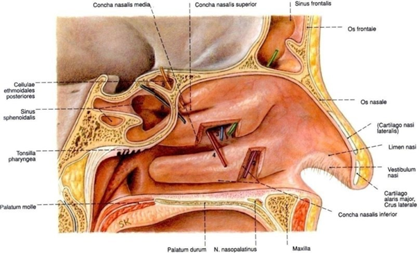
Picture 1. Anatomy Cavity Nose 1
The inferior part of the nasal cavity borders the oral cavity and is separated by palate hard. To direction posterior relate with nasopharynx through the choana. Laterally and in front it is bordered by the external nose. Laterally and behind it is bordered by the orbit of the maxillary sinus, ethmoidal sinus, pterygopalatine fossa, pterygoid fossa. 2,3
Base nose
The floor of the nose is formed by the palatine processes of the maxilla and the horizontal processes of the palatine bone. The roof of the nose consists of the superior and inferior lateral cartilages, And bones OS nasal, OS frontal laminate cribrosa, OS etmoidale, and corpus OS sphenoid. Wall medial cavity nose is septum septum. The septum consists of the septal cartilage, the perpendicular lamina of the ethmoid bone, and the vomer bone. Meanwhile, in the apex of the nose, the septum is completed by skin, subcutaneous tissue, and the greater alar cartilage. 2.3
Lateral wall
Wall lateral can shared become three part, that is in Anteriorly there is the frontal process of the maxillary bone, medially there is the ethmoid bone, maxillary bone and concha, and posteriorly there is the perpendicular lamina of the palatal bone and the medial pterygoid lamina. The most important part of the wall Laterally there are four conchae. The largest and lowest located concha are the inferior concha then the smaller concha are the middle concha, concha superior And Which most small is concha supremacy. Konka supremacy usually will experience rudimentary. 15 Between conchae and the lateral wall of the nose has a narrow cavity called the meatus. There are three meati, namely the inferior, middle and superior meatus. 2,3 Meatus superior or fissure ethmoid is a gap Which narrow between the septum and the lateral mass of the ethmoid bone above the middle concha. The sphenoethmoidal recess located in posterosuperior concha superior And in front concha os sphenoid. The sphenoethmoidal recess is where the sphenoid sinus drains. 2,3
The middle meatus is one of the gaps in which there are the openings of the maxillary sinus, frontal sinus and the anterior part of the ethmoid sinus. Behind the anterior part of the middle concha which is hanging, on its lateral wall there is a crescent-shaped gap called the infundibulum. The opening or crescent-shaped fissure that connects the middle meatus to the infundibulum is called the hiatus semilunaris. The inferior and medial walls of the infundibulum form a drawer-shaped protrusion known as the uncinate process. The ostia of the frontal sinus, the maxillary antrum, and the anterior ethmoid cells open into the infundibulum. The frontal sinus and cells ethmoid front usually culminate in part front above, and sine maxilla culminate in posterior estuary sine frontal. 2.3 Meatus rice inferior is the biggest in between The third meatus has a nasolacrimal duct opening which is approximately 3 to 3.5 cm behind the posterior border of the nostril. 2.3
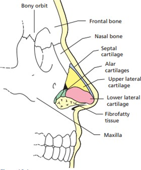
Picture 2. External nose structure 3
Septum Nose
Septum share cavity rice become room right And left. The posterior part is formed by the perpendicular lamina of the ethmoid bone, the anterior part by the septal cartilage, premaxilla and membranous columella. Posteriorly and inferiorly by the vomer bone, maxillary crest, palatine crest and sphenoid crest. 2.3
At the front of the septum there is an anastomosis of the branches of the sphenopalatine artery, anterior ethmoid artery, superior labial artery, and greater palatine artery. called Plexus Kiesselbach ( Little's area ). Plexus Kiesselbach It is superficial and easily injured by trauma, so it is often a source of epistaxis (nosebleeds), especially in children. 2,3
The veins of the nose have the same name and run side by side with the arteries. The veins in the vestibule and the outer structures of the nose drain into the ophthalmic vein which communicates with the cavernous sinus. The veins in the nose do not have valves, which predisposes to easy spread of infection to the intracranial. 2.3
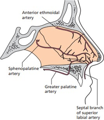
Picture 3. Vascularization nose 3
Part front And on cavity nose get innervation sensory from
n. ethmoidalis anterior, Which is branch from n. nasociliary, Which originates from the ophthalmic n. (N.V1). The rest of the nasal cavity, mostly gets innervation sensory from n. maxilla through ganglion sphenopalatine. Ganglion sphenopalatine ganglion besides providing sensory innervation also provides vasomotor or autonomic innervation to the nasal mucosa. This ganglion receives fibers sensory from n. maxilla (N.V2), fiber parasympathetic from the greater superficial petrosal nerve and sympathetic fibers from the greater petrosal nerve. deep. Ganglion sphenopalatine located in behind And slightly above the posterior end of the middle concha. 3
The olfactory nerve descends from the lamina cribrosa of the undersurface of the olfactory bulb and terminates on the olfactory receptor cells in the olfactory mucosa in the upper third of the nose. 3
In children, there are differences in the size, shape and structure that support the cartilage and bone framework of the nose. The anatomy of the nasal framework in children has a more specific structure than in adults. The healing process of the septal cartilage is an important factor to consider in planning surgical intervention. Children's noses are smaller, shorter and have a larger nasolabial angle. Children also have a flatter tip and a shorter columella. The nasal septum is the main structure which supports the framework of the nose. 3
Growth nose will to be continued after time puberty, on during adolescence the process will end at the age of 18-20 years in men and 16-18 years in women. There are two phases of nose growth, in the first year of life where the endochondral ossification process occurs in the anterior part of the skull base and during puberty the nose grows faster than other periods in life. 3
Physiology Nose
Nose And sine paranasal own a number of function physiological Which important, such as:
Respiratory function: to regulate air conditions (air conditioning ), air filter, humidification and balance in pressure exchange and mechanisms. immunological local. Air inspiration enter to nose going to system respiration through the anterior nares, then up to the level of the middle concha and then down towards the nasopharynx. The air flow in the nose is curved or arched. The inhaled air will experience humidification by hammer mucus. On season hot, air almost fed up by water vapor so that there is little evaporation of inspired air by the mucous membrane, while in winter the opposite will occur. The temperature of the air passing through the nose is regulated so that it is around 37 degrees Celsius. This temperature regulating function is made possible by the many blood vessels under the epithelium and the presence of a wide surface of the concha and septum. Dust particles, viruses, bacteria and fungi inhaled with the air will be filtered in the nose by hairs (vibrissae) in the nasal vestibule, cilia and mucous membranes.
Olfactory function: the true olfactory organs are located in the upper part of the nasal septum and the lateral walls of the nose on each side. The olfactory mucosa contains receptor cells whose axons form the olfactory fila that lead to the olfactory center in the central nervous system. These sensory cells capture the sensations smell And depends on supply air. When there is blockage, the function of the olfactory organ is also greatly hampered which is Clinically, it usually also causes taste disturbances.
Phonetic function: the shape and function of the nose shape each person's characteristics. Nose including in component rigid channel Which to form voice and articulation. If the nose is blocked, there is a lot of secretion and other disorders, the quality of the voice will change and the voice will become nasal.
Nasal reflex: the nasal mucosa is a reflex receptor related to the digestive, cardiovascular and respiratory tracts. Irritation of the nasal mucosa will cause a sneeze reflex and stop breathing. Certain odor stimuli will cause secretion of the salivary glands, stomach and pancreas.
Static and mechanical functions: to lighten the load on the head and protect against trauma and heat protection.
Stenosis Vestibule
Definition and Epidemiology
Nasal vestibular stenosis is a condition in which the nasal vestibule, the front part of the nasal cavity, is narrowed or restricted. This condition can range from mild stenosis to complete atresia and can occur unilaterally or bilaterally. This condition is caused by disruption of the nasal vestibular lining with secondary proliferation of granulation tissue and fibers, which can be caused by various factors such as trauma, infection, and iatrogenic injury to the vestibular skin. 4
The diagnosis of nasal vestibular stenosis is based on clinical examination, and patients typically complain of persistent unilateral or bilateral nasal obstruction. The condition can be congenital or acquired, and causes of acquired stenosis include burns, trauma, infection, and iatrogenic injury to the vestibular lining. Surgical correction is usually accomplished by excision of the fibrous tissue, and various techniques have been described, including scar excision and replacement with local flaps, composite or cartilage grafts, with or without stents. The exact prevalence of nasal vestibular stenosis is unknown, but it is estimated to occur in up to 13% of people with chronic nasal obstruction. 4
Etiopathogenesis
Stenosis vestibule can due to by various factor, covering congenital abnormalities, trauma, infection, and iatrogenic injury. The pathophysiology of nasal vestibular stenosis is not fully understood. However, it is thought that the cause involves a combination of factors, including inflammation, scarring, and contracture of the vestibular skin. Inflammation is a common response to injury. In cases of nasal vestibular stenosis, inflammation can damage layer vestibular And cause formation network scar. Scar tissue is often stronger than normal tissue, and can contract, which can cause narrowing of the nasal vestibule. Vestibular skin is skin Which to coat vestibule nose. Skin This can experiencing contractions, Which can cause constriction on vestibule nose.
Narrowing of the nasal vestibule can cause a variety of symptoms, including nasal obstruction, epistaxis, pain or sensitivity in the nose, dryness or crusting in the nose, a feeling of fullness in the nose, and difficulty breathing through the nose. Stenosis vestibular nose congenital is form Which No common of this disorder, but the symptoms it causes can be the same as post-traumatic nasal vestibular stenosis. Congenital forms of nasal vestibular stenosis generally involve narrow bony structures of the nostrils. 2,4
There are several forms of congenital vestibular stenosis. Stenosis accompanied by choanal atresia is one form of congenital vestibular stenosis. Where there is narrowing of the choanae in the posterior nasal cavity causing symptoms of posterior obstruction. Up to 74% of cases of this disorder are associated with other craniofacial disorders such as CHARGE, Crouzon, Treacher Collins, Pfeiffer, Apert, Antley-Bixler, Marshall-Smith, Schinzel-Giedion syndromes, or cleft lip/palate. In addition, another form of congenital abnormality is congenital piriformis nostril stenosis which is Wrong One condition stenosis vestibule Which can happen on baby which is marked with constriction hole nose piriformis. Deformity This caused by fusion premature And growth excessive on process medial nose maxilla about four months in fetal development. Anterior rhinoscopy shows narrowing of the anterior nasal valve canal, usually affecting the second hole nose in a way bilateral. Stenosis midnasal Congenital is another form of congenital vestibular stenosis disorder which is abnormality Which happen consequence growth excessive bilateral in middle of the nasal cavity, caused by unbalanced growth of the lateral walls of the nose or excessive folding of the nasal septum. 4
Apart from being caused by congenital abnormalities, vestibular stenosis is also often caused by... due to by trauma nose. Formation network scar And contracture after trauma can cause stenosis vestibule rice with obstruction air flow that occurs. Trauma to the alanation or vestibular lining usually occurs due to complex wounds or lacerations. The caudal and lateral parts of the alanation are composed of tissue fibrofatty or fibromuscular which is very easy injury And Contractures occur. Direct trauma to the alanation such as burns and chemical wounds cause tissue damage and cause Scar tissue contracture that causes the alanation to become flat. As a result of tissue damage and scar tissue contracture, negative pressure arises as inspiration further causes the injured vestibule to become increasingly contracted. 3
Other causes of vestibular stenosis include infections and inflammation including chicken pox, leprosy, syphilis, which in the healing process cause scar tissue, resulting in vestibular stenosis. 3Iatrogenic causes of nasal vestibular stenosis refer to causes that due to by intervention or treatment medical. Matter This can includes: 5
Surgical procedures: Several surgical procedures are performed in the vestibule. rice, like septoplasty (correct septum nose Which bent) or rhinoplasty (reshaping of the nose), may inadvertently cause scarring and narrowing of the nasal vestibule, resulting in stenosis.5
Nasal trauma: In some cases, trauma to the nasal vestibule is caused by procedure medical, like use packing nose, Nasal intubation, or the use of nasal instruments, can cause tissue damage and subsequent stenosis. 5
Therapy radiation: Therapy radiation Which targeted on area nose, especially for the treatment of head and neck cancer, can cause tissue fibrosis and scar tissue formation, resulting in nasal vestibule stenosis.5
Diagnosis
The diagnosis of vestibular stenosis can be made based on anamnesis and examination. physical and supporting investigation. The diagnosis of nasal vestibular stenosis is largely based on clinical assessment, therefore, a proper history and physical examination are essential to ensure proper management. 3
Anamnesis
It is very important to know the patient's age, patient complaints such as a runny nose, clogged as well as existence history trauma and also operation nose previously. Patients with nasal vestibular stenosis often complain of unilateral nasal obstruction. or bilateral Which in progress continously. Complaint nose Blockage will be worse if the patient suffers from rhinitis so that the patient has to breathe through the mouth and can cause snoring symptoms. 3
Inspection physique
Physical examination of patients with vestibular stenosis aims to confirm the diagnosis and evaluate case management. Inspection is performed on the outside and inside of the nose to look for changes in shape, deviation, or abnormalities. In patients with vestibular stenosis due to nasal trauma, bimanual palpation examination can be performed. This examination aims to evaluate the presence of crepitation, abnormal nasal bone curvature, and bone irregularities. 3
Inspection rhinoscopy front And endoscopy done For determine the location and severity of stenosis. Anterior rhinoscopy examination should be performed with lighting Which adequate and with patient Which cooperative. Use of a nasal speculum and headlamp may help to expand the view. Vestibular stenosis can occur in various parts of the nose, including the floor of the nose. nose, roof, and lateral walls of the vestibule. On a number of case, stenosis vestibule can accompanied by with damage on concha nose. Besides The stenosis can also involve the entire nasal cavity. 3
The spectrum of nasal vestibular stenosis can range from mild stenosis or narrowing to complete atresia. The degree of nasal vestibular stenosis can be categorized as mild (10%–20% narrowing), moderate (20%–50% narrowing), and severe (>50%), which is determined based on physical examination. According to Johari et al, this classification is not commonly used, but it can provide a rough assessment of the condition and help in describing the degree of stenosis present. 6
Inspection Support
Inspection support can used For know the extent of the damage that has occurred, the location of the stenosis and also the complications that may arise consequence stenosis vestibule. Inspection support The recommended CT-Scan. CT-Scan can be used to confirm the diagnosis of vestibular stenosis after a previous clinical examination or if complications are suspected. 3,6
Management
The goal of treating vestibular stenosis is to restore patency. nasal airway and restore the shape of the nose. Although there are several actions taken to treat vestibular stenosis, treatment to case This can experience difficulty Because causes scarring and has a high recurrence rate. Management of nasal vestibular stenosis is by surgery. The choice of surgical procedure depends on location of stenosis, thickness of obstructed tissue wall, condition of surrounding tissue, degree of external deformity, and condition of alanation. 3
Nasal vestibular stenosis is a challenge for the surgeon due to the lack of structural support from the alar cartilage, the tendency for scar contracture, and the risk of recurrent stenosis. Several surgical options have been described as a one-stage or two-stage repair with various types of grafts and stents. The general approach described in the literature is removal of scar tissue, replacement with a tissue graft, and post-procedure stenting to reduce the risk of recurrent stenosis. Commonly used graft types include cartilage, composite conchal cup graft, composite auricular graft, and mucosal graft with cartilage. Commonly used stent types include metal and thermoplastic, with thermoplastic stents having been described to have better patient comfort with better cosmetic results. more Good compared to with stent metal. Besides various type tissue replacement and stent placement, patients can undergo a single-stage or staged surgical approach. In a retrospective study by Daines et al examined patients who underwent a one-stage or two-stage repair based on the anatomic involvement of the stenosis. Patients with nasal stenosis secondary to isolated ala injury underwent a one-stage repair with conchal cartilage graft and thermoplastic stent. Patients with intravestibular scarring underwent a two-stage procedure, with an additional procedure of scar excision and full-thickness skin graft to create a neovestibular layer, followed by with installation stents. After operation, No There is case stenosis recurrence that occurred within 11 months of follow-up, with a duration of stent placement of less than 2 weeks. 6,7
One alternative to tissue graft placement is flap formation, including intravestibular Z-plasty, triplanar W-plasty, perialar transposition flap, and triple flap. The Z-plasty technique was first described by Horner in 1837, in which the lateral skin excess is transversely moved to lengthen the area along a tight scar line, with the resulting increase in length eliminating contracture. The triplanar W-plasty method allows the transformation of scar tissue into multiple interlocking flaps. The perialar transposition flap involves the nasal floor with lateralization of the nasal base. Bozkurt et al in their study successfully performed a triple flap that included flap transposition shaped boomerang with flap rotation nasolabial and vestibular. 6,8,9
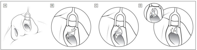
Figure 4. Intranasal Z-plasty procedure. A. The incision is made such that the central incision is an intercartilaginous incision, and the side incisions are directed so that the anterior flap will be based medially and the posterior flap will be based laterally. B, The triangle flaps A and B are then raised. Note the caudal border of the superior lateral cartilage located beneath it. C, The caudal border of the superior lateral cartilage lateral on (area Which shaded) Then lifted For give room for mobilization flaps A and B. D, The flaps are then interdigitated and sutured into position.9
To counteract the continued inspiratory forces that may cause recurrent stenosis after surgical treatment, a vestibular stent is required to provide an opposing force. In the immediate postoperative period, this can be achieved by using an intranasal dressing applied with hydrocortisone, oxytetracycline or polymyxin B sulfate to prevent infection until the surgical wound has healed. 8
Installation stent vestibular Which prolonged required For to prevent collapse of the nasal cavity due to continued inspiratory forces. Various periods of stent use have been suggested in the literature depending on the degree of narrowing and the age of the patient, ranging from six weeks to one year. Minimally invasive methods for progressive nasal dilatation involve balloon dilatation. Some study has been successfully show success with dilator the balloon that give style radial stable compared to with style linear with rigid dilator. Since recurrent stenosis is based not only on constant suction force but also on decreased scar formation and contraction caused by fibroblasts, inhibition of these processes can be achieved by using mitomycin to reduce recurrent stenosis after surgical treatment. Mitomycin C is an antineoplastic antibiotic agent isolated from Streptomyces caespitosus . It works as an alkalizing agent by selectively inhibiting DNA synthesis and DNA cross-linking . At high concentrations it can also press synthesis RNA And protein. Mitomycin C known inhibits fibroblast proliferation and prevents collagen synthesis, which is one of the preventive measures used in conjunction with stent placement. 10
The use of vestibular devices in the postoperative management of vestibular stenosis has been described in the literature to prevent recurrent stenosis, including the use of nasopharyngeal airway tubes, acrylic nasal stents artificial special, And tube suck silicone. Tools This is stent intranasal which must be used by the patient for a certain period of time to ensure results. Which profitable. Stents This has show prevention satisfactory recurrent stenosis. However, they are not directly available for this situation. in where tool the No in accordance Because swelling postoperative, or tool Which too big or damaged, Which can bother period postoperative patient rehabilitation. 6
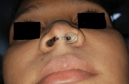
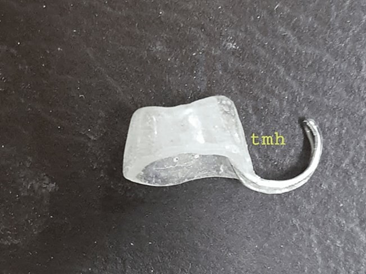
Picture 5. Use stent prosthesis acrylic 6
In addition, vestibular stenosis has a high recurrence rate after surgical repair due to contracture during wound healing. Postoperative scar contracture can occur at any surgical site due to a series of events that occur in wound healing and maturation. Excessive scar contracture can be caused by inherent problems in the physiological process of wound healing, including abnormalities in cell migration and proliferation, synthesis and secretion of extracellular matrix proteins and inflammatory cytokines, as well as injury Which triggered by process remodeling. Besides That, The inward movement of the intact edges of the injured tissue that occurs during wound healing also increases the tendency for the injured vestibule to contract, causing stenosis. This is further aggravated by the constant negative pressure produced by the inspiratory force on the nostrils. 6
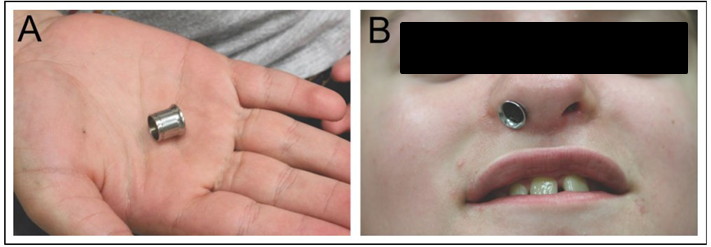
Picture 6. Use stent made from metal
Complications
The most common complication of corrective action of nasal vestibular stenosis is failure to relieve the obstruction. This is usually caused because the surgical technique used is not adjusted to the form anatomy nose patient. Columella Which thick And hole nose Which very small can cause failure, so that synechia occurs even restenosis If No assisted with usage stent. Other complications include epistaxis, infection, scarring, contracture and nasal deformity. become No symmetrical. On area donor Also sometimes become a problem like healing wound Which No perfect, And the emergence keloid. 10
DISCUSSION
Nasal vestibular stenosis is a condition in which the nasal vestibule, the anterior portion of the nasal cavity, is narrowed or restricted. The condition can range from mild stenosis to complete atresia and can occur unilaterally or bilaterally. The condition is caused by disruption of the nasal vestibular lining with secondary proliferation of granulation tissue and fibers, which can be caused by a variety of factors such as trauma, infection, and iatrogenic injury to the vestibular skin. According to Johari et al, nasal vestibular stenosis can be categorized as mild (10%–20% narrowing), moderate (20%–50% narrowing), and severe (>50%), which is determined based on physical examination. This classification is not commonly used, but it can provide a rough assessment of the condition and help in describing the degree of stenosis present. The exact prevalence of nasal vestibular stenosis is unknown, but it is estimated to occur in up to 13% of people with chronic nasal obstruction. 4.6
Vestibular stenosis can be caused by various factors, including abnormalities default born, trauma, infection, And injury iatrogenic. Pathophysiology Nasal vestibular stenosis is not fully understood. However, it is thought that the cause involves a combination of factors, including inflammation, tissue formation, scar, and contractures on vestibular skin. The diagnosis of vestibular stenosis can be established based on anamnesis, physical examination and supporting examinations. The diagnosis of nasal vestibular stenosis is largely based on clinical assessment, therefore, a proper history and physical examination are essential to ensure proper management. 3
The goal of treating vestibular stenosis is to restore nasal airway patency and restore nasal shape. Treatment for nasal vestibular stenosis is surgical. The choice of surgical procedure depends on the location of the stenosis, the thickness of the obstructed tissue wall, the condition of the surrounding tissue, the degree of external deformity, and the condition of the alanation. Common approaches described in the literature include removal of scar tissue, replacement with graft tissue, and post-procedure stenting to reduce risk stenosis repetitive. In matter This stent functioning to keep the vestibule always expanded or to act as a support. Stents It can also be used temporarily while waiting for surgical procedures, especially for incomplete stenosis and congenital abnormalities. 7
One alternative for tissue graft placement is flap formation, including intravestibular Z-plasty, triplanar W-plasty, perialar transposition flap, and triple flap. Bozkurt et al in their study successfully performed a triple flap that included flap transposition shaped boomerang with flap rotation nasolabial and vestibular. According to Daines et al, triplanar W-plasti, Which involving removal of scar tissue from within the nostrils and the formation of several flaps, which then interdigitate to provide epithelial coverage. Use of stents acrylic during 5 month after W-plasti recommended For prevent restenosis. In studies Which done Choudhury et al use technique Z-plasty simple and obtained very satisfactory results, showing statistically significant improvement in deformity. 7,8,11
Commonly used stent types include metal and thermoplastic, where thermoplastic stents have been described to have better patient comfort with better cosmetic results compared to metal stents. In addition to various type replacement network And installation stents. On a retrospective study that done Daines et al, patient with stenosis nose secondary consequence injury isolated undergo repair One stage with graft cartilage konkal And thermoplastic stent. In patients who have intravestibular scar tissue, a two-stage procedure is performed, with an additional procedure in the form of scar tissue excision and grafting. skin thickness full For create layer neovestibular, followed with stent placement. After surgery, no cases of recurrent stenosis occurred within 11 months of follow-up, with a duration of stent placement of less than 2 weeks. 6,7
The use of vestibular devices in the postoperative management of vestibular stenosis has been described in the literature to prevent recurrent stenosis, including the use of nasopharyngeal airway tubes, acrylic nasal stents artificial special, And tube suck silicone. Tools This is stent intranasal which must used by patient in term time certain For ensure favorable outcome. In a case report published by Shankar et al a custom-fit acrylic prosthesis stent was used in the patient's nose where use circle wire as buffer. Where stent used for a period of three months, then in the follow-up in the sixth month, good results were still obtained. Pradhan et al in his case report used a stent made specifically from an endotracheal tube (No. 8) and was followed up for 6 months, obtained adequate nasal patency and good aesthetics. In addition That, Johari et al try use glue gun stick (glue candle shoot) in patients with left vestibular stenosis caused by traffic accidents. In these patients, dilation of the left nostril was performed using a Hegar dilator and resection of fibrous tissue using the jumping-man Z-plasty technique. A week after operation, glue candle shoot This used For dilation routine. 2% lidocaine gel was used to lubricate and facilitate stick placement and to prevent friction that could potentially injure the nasal mucosa. Continuous daily dilation of the nasal vestibular opening was recommended for three consecutive months. At 3-month follow-up , the patient was able to easily insert a standard-sized 7 mm diameter glue gun stick into both nostrils. The patient also had significant improvement in her nasal obstruction and had no recurrence of nasal stenosis. Use of glue candle shoot This can become alternative Because access Which easy, available, and cost-effective, and is based on the principle that in vestibular stenosis, the need for continuous dilation of the nasal vestibule in the postoperative period is essential to properly form the tissue around the narrowed area. 12,13
CONCLUSION
Nasal vestibular stenosis is a condition in which the front part of the nasal cavity, Which called vestibule nose, experience constriction or restrictions. Level severity stenosis This Can light until cause blockage total, and can happen on One side nose (unilateral) or second side nose (bilateral). Condition This due to by disturbance on layer in vestibule nose which experiences excessive growth of granulation tissue and fibers, which can be caused by trauma, infection, or injury that occurs during a medical procedure on the skin of the nasal vestibule.
Accurate diagnosis and appropriate treatment are essential in management. stenosis vestibule nose. Prevalence Certain condition This No It is not known for certain, but is thought to occur in some people with chronic nasal obstruction. CT scans can be used to confirm the diagnosis of vestibular stenosis. after done inspection clinical previously and also when suspected complications arising.
Treatment of nasal vestibular stenosis aims to restore patency. road breath nose as well as return form nose. Approach Commonly used are scar tissue removal, replacement with graft tissue, and post-procedure stent placement to reduce the risk of recurrent stenosis. The choice of surgical procedure depends on the location of the stenosis, the thickness of the obstructed tissue wall, the condition of the surrounding tissue, the degree of external deformity, and the condition of the ala nasi.
REFERENCE
Soetjipto D, Mangunkusumo E, Wardani RS. Nasal obstruction. In: Soepardi EA, Alexander N, Bashiruddin J, Restuti RD, editor. Textbook knowledge health ear, nose, throat, head And neck. Edition to-
7. Jakarta: FKUI; 2012:96- 100
Hsu DW, Suh JD. Anatomy and physiology of nasal obstruction. Otolaryngologic Clinics of North America. 2018;51(5):853–65
Corbridge R. Essential ENT. 2nd ed. Hoboken: CRC Press; 2011.
Galluzzi F, Garavello W, Dalfino G, Castelnuovo P, Turri-Zanoni M. Congenital bony nasal cavity stenosis: A review of current trends in diagnosis and treatment. International Journal of Pediatric Otorhinolaryngology.
Ebrahimi A, Shams A. Severe iatrogenic nostril stenosis. Indian J Plast Surg. 2015;48:305-8.
Johari SNA, Mat Saad AZ, Ramli RR, Shukri NM. European Journal of Medical Case Reports. Post-corrective nasal stenosis patency rehabilitation using glue gun stick: a case report on the novel management strategy. 2020 May 4;4(5):218–22.
Daines SM, Hamilton GS, Mobley SR. A graded approach to repairing the stenotic nasal vestibule. Archives of Facial Plastic Surgery. 2010;12(5).
Bozkurt M, Kapi E, Kuvat SV, Selçuk CT. Repair of nostril stenosis using a triple flap combination: Boomerang, nasolabial, and vestibular rotation flaps. The Cleft Palate-Craniofacial Journal. 2012;49(6):753–8
Dutton JM, Schneider JS, Ray CA. The intranasal Z-plasty for nasal valve collapse. Advanced Aesthetic Rhinoplasty. 2013;367–72.
Van Schijndel O, van Heerbeek N, Ingels KJAO. Current treatment of nasal vestibular stenosis with CO2-laser surgery; Prolonged vestibular stenting versus intraoperative mitomycin application. A case series of 3 patients. International Journal of Pediatric Otorhinolaryngology. 2014;78(12):2308–11.
Choudhury N, Hariri A, Saleh H. Z-plasty of the alarm subunit to correct nasal
vestibular stenosis. Otolaryngology–Head and Neck Surgery. 2014;150(4):703–6.
Shankar R, Dubey M. Custom nasal stent fabrication for post-traumatic nasal obstruction - a case report. Cureus. 2022
Pradhan P, Preetam C. Columellar strut in correcting the recurrent vestibular stenosis. Indian Journal of Otolaryngology and Head & Neck Surgery. 2021;74(S2):1430–2.
asfsaf
Comments