SYNDROME COLLET SICARD AS COMPLICATIONS FROM MALIGNANT OTITIS EXTERNA
I Made Nudi Arthana
Although the cause of the infection and inflammation Less commonly, cases of polyarteritis nodosa, Trousseau syndrome, otitis media, Lyme disease, and varicella-zoster causing Collet-Sicard syndrome have been reported. 3 There have been only 2 reported cases of osteomyelitis or inflammatory ear disease causing Collet-Sicard syndrome before our case. 1
LITERATURE REVIEW
Definition Syndrome Collet-Sicard And Otitis External Malignant
Definition And Anatomy Syndrome Collet- Sicard
CSS, first described by Collet and Sicard, is a rare condition. Which involving paralysis combination And unilateral on nerve cranial IX, X, XI, and XII. This syndrome usually develops gradually and its clinical presentation may be complex, therefore the diagnosis is often delayed. Sometimes this syndrome occurs in an incomplete form, resulting in another type of syndrome known as jugular foramen syndrome. When CSS occurs together with ipsilateral Horner syndrome, it is called Villaret syndrome. 7,8
When syndrome Collet-Sicard suspected as process focal on base skull, then primary neoplasm or metastasis must be ruled out but also non-neoplastic disorders such as vascular disease especially dissection or aneurysm of the internal carotid artery and thrombosis of the sigmoid-jugular complex. 3 CSS can be part of head trauma 9 diseases Lyme, neuropathy of the nerves cranial idiopathic, And polyarthritis nodosa.3 This matter can caused by iatrogenic causes such as cardiac surgery and endovascular procedures. 10 To date, only two cases of CSS associated with ear infection have been published. 1
The CSS is closely related to the course of the lower four cranial nerves through the jugular foramen and along the major blood vessels of the neck (Figure 1).

Picture 1. Journey four nerve cranial part lower through foramen jugular and along the major blood vessels of the neck. 11
The jugular foramen is located just above and to the side of the occipital condyle. It is divided into three distinct compartments by the temporal intrajugular process which continues with two septum Dura intrajugular material thick. Anteromedial part, which in a way historical known as pars nervous, is track For CN IX together with inferior petrosal sinus. The middle portion, which consists of two dural septa, is referred to as the inferior petrosal compartment. intrajugular And containing CN X And CN XI, whereas artery pharyngeal The ascending artery passes between the two to supply the posterior meningeal artery. The intrajugular compartment this is in a way anatomical including in the posterolateral sigmoid foramen jugular vein, historically known as the pars vascularis and is the portal of entry for the jugular bulb, Which is corner the protrusion between sine sigmoid and veins internal jugular (IJV). 12,10
During his journey in outside foramen jugular, CN IX moored to vessels the main blood supply to the neck by dense connective tissue and is thus very susceptible to injury. vessels blood. 11,12 Accessory nerve cranial separate self from the nerves spinal accessory nerve which then rejoins the middle ganglion of the vagus nerve, which eventually becomes the recurrent laryngeal nerve. Finally, the hypoglossal canal passes through the occipital condyle. CN XII exits the hypoglossal canal medially into the jugular foramen And walk in between IJV And artery carotid internal (ICA), before cross over to direction lateral And going to to front. By Because That, fourth nerve cranial part the lower and main blood vessels of the neck are closely interconnected. 13
Definition And Epidemiology Otitis Malignant External
Malignant otitis externa (OEM) is an aggressive, but rarely life-threatening, ear infection. It is an infection of the soft tissues of the external ear and surrounding structures, which spreads to involve the periosteum and skull base. The first case of malignant otitis externa was reported in 1838 by Toulmouche and the term “otitis externa” was coined. malignant" First time used by Chandler on in 1968, because its high morbidity and mortality before the introduction of appropriate antibiotic treatment 14 . Infection This part big seen on patient Which experience disturbance immunocompromised individuals over 65 years of age, especially those with diabetes, hematologic disorders (eg, leukemia or granulocytopenia) or arteriosclerosis. 15 The most common causative agent is Pseudomonas aeruginosa, but other bacterial species, including Staphylo-coccus Methicillin-resistant aureus (MRSA), as well as fungal species have been reported. Symptom Which general appear including otalgia, otorrhea, flavor full in ear and hearing loss. 16 The most common complications are cranial nerve palsy, osteomyelitis, especially of the facial nerve, meningitis, and brain abscess. 17
The annual incidence of malignant otitis externa in the age group under 18 years is the lowest, while the age group over 65 years is the highest. Older adults are susceptible to OEM, due to the higher frequency of immunocompromising diseases, such as diabetes mellitus and chronic kidney disease. Interestingly, it has been found that 27.8% of OEM cases are comorbid with hypertension, Which in line with report other, but need investigation more carry on. Until moment This, only There is series case not enough from 80 case Which show low OEM incidence. 18,19
Etiology
Ethology Syndrome Collet- Sicard
CSS can triggered by a number of condition, for example, process invasive in base skull (primary or metastatic tumor), trauma, vascular lesions, inflammatory processes, and iatrogenic complications. The most commonly affected cranial nerve is cranial nerve VIII, followed by cranial nerves V, IX, X, and XI. Pure motor cranial nerves are less commonly affected. 20 Glomus tumor, or paraganglioma, is tumor with vascularization tall Which consists of from cells that originate from the neural crest during embryonic development. While 90% of paragangliomas arise in the adrenal glands, only 3% develop in the head and neck. 21 Tumor This grow in near foramen jugular And can expand to room intracranial and extracranial. Surgical resection is the treatment of choice for neurinomas and paragangliomas. However, stereotactic radiosurgery is the main treatment alternative for patient age carry on, patient with condition clinical Which bad, or patient Which have residual or recurrent lesions after surgery. 22
Search literature about CSS Which available in PubMed show total 51 cases published between 1915 and 2012. Table 1 summarizes the various etiologies of CSS. Some literature from PubMed related to the onset of CSS between 1915 and 2012 2020. Term search “syndrome Collet-Sicard” And “Collet Sicard” used, and 100 reports were found. This syndrome can be caused by a variety of conditions, including tumors, vascular lesions such as the carotid artery or jugular vein, trauma, iatrogenic, and inflammation. With the three most commonly cited causes being invasive metastatic disease, carotid artery dissection, and skull base fracture. Summary of known cases with etiology syndrome Collet-Sicard Which reported in literature since 1915 has been included as a reference, adapted from the excellent review by Gutiérrez Ríos et al and updated to reflect cases since 2013.
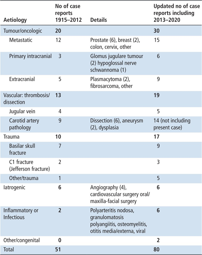
Among the cases reported in 100 publications, 17 (33.33%) patients experienced CSS due to trauma factors, with head trauma and Jefferson fractures being the dominant causes. CSS due to vascular factors accounted for 19 (37.25%) patients, with internal carotid artery disorders being the most common cause. A total of 30 cases of CSS due to tumor factors accounted for 58.82% of the total number of cases, 15 of which were due to malignant tumor metastasis, accounting for 50% of CSS caused by tumors. CSS due to prostate cancer had the highest reporting frequency. Laigle-Donadey et al. stated that the most common skull base metastases were prostate and breast cancers. Rhiannon et al. performed a total of 5,644 imaging examinations, including the brain, in 4,341 Australians with prostate cancer. 27 Eight from patient This diagnosed with metastasis intracranial simultaneously, Which
show low incident metastasis intracranial on patient cancer prostate (0.18%). In recent years, research on the incidence of intracranial metastasis from prostate cancer has been rare, and there is little evidence to suggest that prostate cancer is associated with an increased risk of intracranial metastatic complications.
A total of 16 cases of CSS were caused by primary benign intracranial tumors, accounting for 37.2% of cases caused by tumors; two of these cases were caused by paragangliomas, 28,29 accounting for 12.5% of cases caused by benign tumors. The cases reported in this article fall into the category of benign tumor etiology. Invasive tumor metastasis, vascular lesions such as internal carotid artery obstruction, and head trauma are common causes of CSS; the number of cases arising from primary intracranial tumors is relatively small. 30
Etiology And Factor Risk Otitis External Malignant
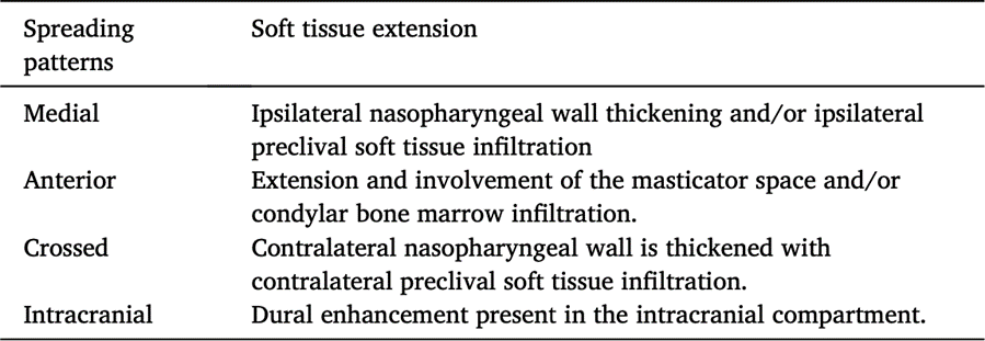
The most common pathogen causing OEM is Pseudomonas aeruginosa of all OEM cases (50-90%). Followed by other pathogens such as Proteus mirabilis, Aspergillus fumigatus, Proteus spp, Klebsiella spp, And Staphylococcus Also has reported. 19 Index suspicion Which tall For organism atypical, like MRSA, must maintained in patients with signs and symptoms of OEM who do not have diabetes. 31 OEM usually begins in the external auditory canal (ECA) and spreads to the stylomastoid foramen and then to the mastoid tip and jugular foramen. Eventually, the septic process extends to the apex. petrous And fossa cranial middle. Kwon et al., describe four pattern soft tissue extension spread: medial, anterior, transverse, and intracranial spread, which are best described in Table 2. One of the severe complications that can arise from OEM is the involvement of cranial nerves, especially the facial nerve. 16
Table 2. Distribution Extension Network Soft on Otitis External Malignant
The most common risk factor reported in the literature for the development of OEM is diabetes mellitus, with an estimated 90-100% of patients with OEM having DM. Diabetes mellitus has been suggested to predispose patients to OEM by inducing microangiopathy in channel ear And hinder chemotaxis cell blood the white one result in improvement vulnerability to infection. 31 Factor risk main others are immune suppression, such as patients suffering from Human Immunodeficiency Virus (HIV), transplant patients, or patients with advanced cancer. OEM must always suspected when patient with immunosuppression appear with symptoms of otitis externa, especially if the otitis does not respond to the usual therapies. Some study has find that age carry on is factor important others. Almost 20% the patient does not have history medical previously. On year 2019, Bruschini et al. report a patient Which No like most subject Which caught impact, no suffer diabetes or disturbance immunity body, but previously Once treated with radiotherapy to the head and neck region 20 years before presentation of OEM. Radiotherapy can induce process necrosis which is very slow on the bones And Bacterial infections can invade necrotic tissue. 32
Manifestation Clinical
Syndrome Collet- Sicard
CSS manifest with symptom clinical Which can recognized:
CN IX palsy causes anesthesia of the soft palate and loss of the gag reflex.
CN X palsy causes paralysis of the vocal cords, usually trapped in a medial position.
CN XI paralysis manifests as drooping shoulders indicating trapezius muscle paralysis and difficulty turning the head to the contralateral side indicating sternocleidomastoid muscle paralysis.
CN XII palsy identified with downsizing tongue ipsilateral ( ipsilateral protraction). 13
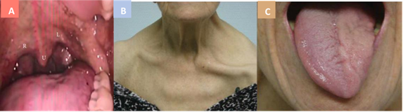
Figure 2 Collet-Sicard syndrome: (A) Elevation disturbance on the right side of the soft palate. compared to with side left with refers to uvula (U) (CNX palsy right). (B) paralysis nerve cranial XI left which result in drooping on shoulder and clavicular prominence; (C) left cranial nerve XII paralysis causing atrophy on the left side tongue 13
The cases were classified into two groups: “CSS”, referring to patients exclusively showing involvement of nerves IX-XII, and ‘CSS-plus’, which corresponded to cases with CSS and other neurological disorders. The researchers included 135 patients from 126 articles, of which 84 (67.7%) were male. The most commonly reported clinical manifestations were dysphagia and dysphonia. Etiology Which most general is tumor on 53 case (39.6%) And vascular on 37 cases (27.6%). The majority of patients showed partial or complete improvement, with more than half receiving conservative treatment. The most frequently affected anatomy is the foramen jugular (44.4%) And room retrostyloid parapharyngeal (28.9%). Around 21% patients had other CN disorders, with the seventh and eighth CNs being the most frequently affected. In conclusion, although there is a need for greater precision in reporting CSS, this syndrome has clear utility in identifying the localization of jugular foramen and parapharyngeal retrostyloid space pathology. 33
Otitis External Malignant
This condition often presents as intractable otalgia, which is often the more getting worse, And spread to face side ipsilateral. 34 Manifestation clinical from The disease persists for more than a month, including chronic otorrhea, headache, and cranial nerve involvement. Singh et al. showed in 2018 that nocturnal pain was the most common feature, followed by ear discharge , hearing loss, and temporal-romandibular joint pain. Otoscopy may reveal edema KAE And existence network granulation or polyp on inferior portion of the EAC near the junction of the osseous and cartilaginous portions. 35 Cranial nerve palsies at presentation have been reported and are well recognized as complications, with the facial nerve being most commonly involved due to its proximity to the external auditory canal. This factor increases the mortality rate by 50%. Undiagnosed OEM or treated in a way partial can in a way progressive spread to skull base and cause serious complications such as lateral sinus or internal jugular vein thrombosis, meningitis, Bezold's abscess, and cranial nerve palsy.36
Inspection Support
Diagnosis Syndrome Collet- Sicard
Once clinical suspicion of CSS is established, time- and cost-effective imaging studies that allow for an accurate diagnosis should be performed. Given the frequency of tumoral, vascular, and traumatic causes, imaging may include computed tomography of the brain with or without angiography and/or magnetic resonance imaging of the brain with or without angiography. If these paraclinical investigations are not diagnostic, digital subtraction angiography is mandatory. In this review, 60.3% of patients (44/73) underwent computed tomography, 57.5% of patients (42/73) underwent magnetic resonance imaging, 15.1% of patients (11/73) underwent angiography subtraction digital, And 9.6% patient (7/11) undergo tomography positron emission or scintigraphy. 13
Diagnosis Otitis Malignant External
Inflammatory markers, such as erythrocyte sedimentation rate (ESR), white blood cell count, or proteins reactive C (CRP) can increase on patient with OEM. Computed computed tomography (CT) scan has proven to be a useful tool for diagnosis and prognosis prediction. Magnetic resonance imaging (MRI) is useful for soft tissue involvement, but is inadequate for bone involvement, is expensive, and is not available in all centers. Magnetic resonance imaging provides excellent anatomic localization and soft network in arrangement suspected osteomyelitis. Bone changes can detected as early as 3-5 day since the emergence disease. Sensitivity MRI in detection of diabetic osteomyelitis has been reported to be 90%, with a specificity of 79% .37,38
Diagnosis Appeal
Understanding Which appropriate about anatomy nerve cranial, inspection physique Which careful and approach gradually to various process disease Which can cause dysphagia symptoms And dysphonia on patient This, support enforcement end diagnosis. Correctly differentiating objective neurologic examination findings is essential to determining accurate neurologic localization. Considerations of where these findings will be localized include lesions in medulla medial, like consequence stroke ischemic or demyelination, Which influence dorsal vagal and hypoglossal nuclei and the nucleus of the tractus solitarius. This can cause deviation of the tongue contralateral, tachycardia And change voice. Besides That, process meningeal, Good due to infection or inflammation, such as neurosarcoidosis, can selectively affect some nerve cranial in a way unilateral or bilateral. Inspection MRI brain Which normal and profile fluid cerebrospinal Which normal make consideration diagnostic This become smaller the possibility. Disease Lyme can influence nerve cranial individual, with the facial nerve being the most common, however this is less likely given that serology Lyme negative. Tuberculosis Also can influence nerve cranial isolated, although more frequently involving the ocular cranial nerves, as well as a lack of risk factors.
Left tongue atrophy and fasciculations would localize to the left hypoglossal nerve. When combined with left uvular deviation, localized to the right vagal nerve palsy, and concern for facial weakness, this would suggest a lower motor neuron process. The differential would then include a post-viral inflammatory process. or demyelination. For example can covers polyneuropathy demyelination inflammation acute bulbar, in which a normal cerebrospinal fluid profile may be seen initially, or myasthenic syndrome, which is less likely in this patient given its acute onset, the absence of extraocular muscle abnormalities, intermittent diplopia or shortness of breath.39
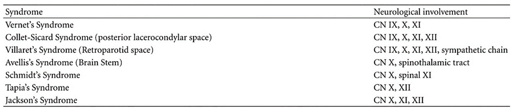
Table 2. Syndrome Jugular Foramen 9
Management
Syndrome Collet- Sicard
CSS is a very rare clinical condition resulting from direct irritation or compression of the lower four CNs during their passage through the jugular foramen or along the neck vessels. Causes may include skull base tumors, vascular injuries, traumatic lesions, infectious diseases, or more anecdotal causes, management of patients with CSS requires a multidisciplinary approach regarding the CN deficit and the patient's general status.
Tumor Base Skull
Treatment of patients with skull base tumors involves evaluation performance status. In cases of metastasis, the Stepwise Prognostic Assessment is a preoperative screening tool aimed at determining candidates for further surgical treatment. 40 Surgical removal of tumors located around the jugular bulb is a procedure Which definitive. Knowledge Which appropriate about anatomy surgery area this is prerequisite For do operation functional, remember proximity to the bulbospinal junction, the presence of the vertebral arteries, and the lower CNS. 41 Beyond the well-known complications, such as intracranial infection and cerebrospinal fluid leak, the surgical procedure itself can worsen CNS deficits. 42 Taking this into account, radiotherapy palliative can become alternative other, moreover Again on tumor Which very aggressive or multimetastatic patients. 43
Lesion Traumatic
Occipital condyle fractures as well as C1 Jefferson fractures mostly require maintenance orthopedics. However thus, fracture Which No consolidated And fracture Which No symptomatic (for example, when There is deficit CN) may indicate the need for surgical treatment. 44
Lesion Vascular
Management dissection artery carotid internal (ICA), aneurysm ICA, And IJV thrombosis supported by guidelines Which has accepted in a way wide. Often time, patient who suffers disease vascular This can cured with management current , which combines blood thinners and sometimes endovascular therapy.45,46
Otitis External Malignant
Before anti-Pseudomonas aeruginosa antibiotics were introduced, mortality rates reached 67% and surgery was considered the mainstay of treatment 37 . The use of oral and topical fluoroquinolones in systemic treatment has allowed treatment take care road Which effective And has reduce need hospitalization. Treatment with a duration of 4-6 weeks may not be adequate for patients at risk tall experience infection repetitive, like patient with infection Because MRSA and end-stage renal disease. 47
Surgery should be considered in aggressive or advanced cases, facial nerve palsy, sterile deep tissue cultures and refractory OEM. Refractory OEM is defined as no clinical improvement after six weeks of conventional therapy. Extensive bone erosions require mastoidectomy. Surgery reduces the local infective burden, removes necrotic tissue and allows for new tissue growth, which increases local vascularization allowing anti biotic systemic achieve area Which needed. 48 Regimen Treatment should be individualized, with multidisciplinary collaboration among specialties, including endocrinology, internal medicine, and infectology.
Prognosis
CSS with etiology infection And vascular show level recovery function The highest CN. First, some of the CN paralysis can be caused by inflammation that can heal over time. Second, cerebral palsy or CN palsy can due to by effect mass abscess base skull or lump intravascular. Shrinking effect mass, blessing maintenance Which appropriate, can show recovery symptoms that progressive. On the contrary, if excision surgery tumor Which located in around foramen The jugular vein is technically more challenging. Most patients undergo subtotal surgical excision. Which followed with radiotherapy, And condition time Which very fragile make are increasingly susceptible to post-operative complications. 49
REFERENCE
Bonda S, Tun KM, Asad S. A Case of Collet-Sicard Syndrome Caused by Otitis Externa. Cureus. 2022;14(7).
Lee M, Heo Y, Kim T. Vernet's Syndrome Associated with Internal Jugular Vein Thrombosis. J stroke Cerebrovasc Dis Off J Natl Stroke Assoc. 2019 Feb;28(2):344– 6.
Handley TPB, Miah MS, Majumdar S, Hussain SSM. Collet-Sicard Syndrome from Thrombosis of the Sigmoid-Jugular Complex: A Case Report and Review of the Literature. Int J Otolaryngol. 2010;2010:1–5.
RD's blessing. Comprehensive management of malignant otitis externa with tuberculosis and cranial nerve paresis in geriatrics. Auto Rhino Laryngol Indonesia. 2020;50(1):77.
Rajputs, S., Arain, A., Cloud, M., Akhtar, S., Adeel M. Malignant otitis external: Cranial nerve palsies, skull-base erosions and prognosis. AKU Institutional Repos J Cranio- Maxillary Dis. 2013;2(2):130–3.
Neo S, Lee TO. Collet-Sicard syndrome: A rare but important presentation of internal jugular vein thrombosis. Pract Neurol. 2017;17(1):63–5.
FJ Collets. Sur un nouveau syndrome paralytique pharyngo-larynge par blessure de guerre (hemiplegic glosso-laryngo-scapulo-pharyngee). Lyon Med. 1915;124:121–9.
Sicard JA. Syndrome du carrefour condylodechire posterieur (type pur de paralysie des quatre derniers nerfs craniens). Marseille Med. 1917;53:385–97.
R. GutiérrezRíos, A. CastrilloSanz, C. GilPolo, MIZamoraGarcía, N. MorollónSánchez- Mateos AM. Collet-Sicard syndrome. Neurology. 2015;30(2):130–4.
Sokhi DS, Mithi CW, Ebrahim FA, Salyani A, Waa S RM. Collet-Sicard syndrome due to concurrent extramedullary intra-cranial plasmacytoma and jugular venous sinus thrombosis in multiple myeloma. Clin Case Rep. 2021;9(7).
Tummala RP, Coscarella E M.J. Surgical anatomy of the jugular foramen. Operation Tech Neurosurg. 2005;8:2–5.
Lin J, Zhou L HD. Internal carotid artery aneurysm presenting US lower cranial nerve palsies. World Neurosurg. 2023;173:23–4.
Shamanna K GV. Changing trends in the management of malignant otitis external: our experience. Otolaryngol Res. 2018;7(1):9–14.
Beucler N. Combined Deficit of the Four Lower Cranial Nerves also Known US theSyndrome of Collet-Sicard: A Systematic Review and Meta-analysis. Asian J Neurosurg. 2024;19(02):112–25.
Sokołowski J, Lachowska M, Karchier E, Bartoszewicz R, Niemczyk K. Skull base osteomyelitis: factors implicating clinical outcome. Acta Neurol Belg [Internet]. 2019;119(3):431–7. Available from: https://doi.org/10.1007/s13760-019-01110-w
Hatch JL, Bauschard MJ, Nguyen SA, Lambert PR, Meyer TA, McRackan TR. Malignant Otitis Externa Outcomes: A Study of the University HealthSystem Consortium Database. Ann Otol Rhinol Laryngol [Internet]. 2018 Jul 2;127(8):514–
WA, Aremu SK, Olatoke F, Olajuyin AO, Ogundipe KO. Epidemiology of otitis externa in developing countries. Int J Recent Sci Res. 2017;8(6):18023–7.
Cheng YF, Yang TH, Wu CS, Kao YW, Shia BC, Lin HC. A population-based time trend study in the incidence of malignant otitis externa. Clin Otolaryngol [Internet]. 2019;44(5):851–5. Available from: https://onlinelibrary.wiley.com/doi/abs/10.1111/coa.13387
Which TH, Xirasagar S, Cheng YF, Wu CS, Kao YW, Shia BC, et a. Malignant Otitis Externa is Associated with Diabetes: A Population-Based Case-Control Study. Ann Otol Rhinol Laryngol [Internet]. 2020 Jan 24;129(6):585–90. Available from: https://doi.org/10.1177/0003489419901139
Sarma S, Sekhar LN SD. Nonvestibular schwannomas of the brain: a 7-year experience. Neurosurgery. Neurosurgery. 2002;50:437—48.
Myssiorek D. Head and neck paragangliomas: An overview. Otolaryngol Clin North Am [Internet]. 2001;34(5):829–36. Available from: https:// www.sciencedirect.com/science/article/pii/S0030666505703492
Jordan JA, Roland PS, McManus C, Weiner RL GC. Stereo-tastic radiosurgery for glomus jugulare tumors. Laryngoscope. 2000;110:35—8.
Paparounas K, Gotsi A, Apostolou F AN. Collet—Sicard syndrome disclosing glomus tumor of the skull base. Euro Neurol. 2003;49:03—5.
Jiménez-Caballero PE, Marsal-Alonso C, Méndez-Cendón JC, Álvarez-Tejerina A. Collet-Sicard syndrome secondary to a glomus tomour: evaluation by means of magnetic resonance. RN. 2004;39(11):1072–3. Available from: https://doi.org/10.1177/000348941877805
Garcia-Escrivà A, Pampliega Pérez A, Martín-Estefania C BC. Schwannoma of the hypoglossal nerve presenting as a syndrome of Collet—Sicard. Neurology. 2005;20:311–3.
Laigle-Donadey F, Taillibert S, Martin-Duverneuil N et al. Skull-base metastases. J Neurooncol. 2005;75:63–69.
McBean R, Tatkovic A WD. Intracranial metastasis from prostate cancer: investigation, incidence, and imaging findings in a large cohort of Australian Men. J Clin Imaging Sci. 11:24.
Mutlu V, Ogul H. Magnetic Resonance Imaging Features of Collet-Sicard Syndrome Associated With Glomus Jugulare Paraganglioma. J Craniofac Surg. 2019 Sep;30(6):e574–6.
Dey JK, Carlson ML. Jugular Paraganglioma Presenting With Collet-Sicard Syndrome. Mayo Clinic Proc. 2019 Sep;94(9):1832–3.
Yanxun Huang, Yanli Huang H.J. Collet–Sicard syndrome caused by a paraganglioma in the region of the jugular foramen on one side: A case report and review. Sage J. 2024;
Byung soo YJ, Patel J, Nguyen SA, Lambert PR. Necrotizing Otitis External: A Systematic Review and Analysis of Changing Trends. Auto Neurotol [Internet]. 2020;41(8). Available from: https://journals.lww.com/otology- neurotology/fulltext/2020/09000/necrotizing_otitis_externa a_systematic_review.2.aspx
Long DA, Koyfman A, Long B. An emergency medicine-focused review of malignancies otitis externa. Am J Emerg Med [Internet]. 2020;38(8):1671–8. Available from: https:// www.sciencedirect.com/science/article/pii/S0735675720303132
Aguilera-Pena MP, Castilbanco MA, Osejo-Arcos V, Aponte Caballero R, Gutierrez-Gomez S, Abaunza-Camacho JF, et al. Collet-Sicard syndrome: a scoping review. Neurosurg Rev [Internet]. 2023;46(1):244. Available from: https://doi.org/10.1007/s10143-023-02145-7
Balakrishnan R, Dalakoti P, Nayak DR, Pujary K, Singh R, Kumar R. Efficacy of HRCT Imaging vs SPECT/CT Scans in the Staging of Malignant External Otitis. Otolaryngol Neck Surg [Internet]. 2019;161(2):336–42. Available from: https://aao- hnsfjournals.onlinelibrary.wiley.com/doi/abs/10.1177/0194599819838834
Croonenburg AMJL, van from See WL, Both of CPR, van Tilburg M, van Tongeren J, Postma AA. Advanced Imaging Techniques in Sku ll Base Osteomyelitis Due to Malignant Otitis Externa. Curr Radiol Rep [Internet]. 2018;6(1):3. Available from: https://doi.org/10.1007/s40134-018-0263-y
Marina S, Goutham MK, Rajeshwary A, Vadisha B, Devika T. A retrospective review of 14 cases malignant otitis externa. J Auto [Internet]. 2019;14(2):63–6. Available from: https:// www.sciencedirect.com/science/article/pii/S167229301830062X
Peled C, Parra A, El-saied S, Kraus M, Kaplan DM. Surgery for necrotizing otitis externa—indications and surgical findings. Eur Arch Oto-Rhino-Laryngology [Internet]. 2020;277(5):1327–34. Available from: https://doi.org/10.1007/s00405-020-05842-x
Peled C, Kraus M, Kaplan D. Diagnosis and treatment of necrotising otitis externa and diabetic foot osteomyelitis – similarities and differences. J Laryngol Otol [Internet]. 2018/08/28. 2018;132(9):775–9. Available from: https:// www.cambridge.org/core/product/F91B62346D5099F1B64FC4B0C612AA7 3
Evan J, Johansen M, Akst LM. Dysphagia, dysphonia and a deviated tongue: Diagnosing Collet-Sicard syndrome. BMJ Case Rep. 2021;14(5):3–6.
Sperduto PW, Case N, Robert D, Xu Z, Shanley R, Luo X, et al. Summary report on the graded prognostic assessment: an accurate and facile diagnosis-specific tool to estimate survival for patients with brain metastases. J Clin Oncol Off J Am Soc Clin Oncol. 2012 Feb;30(4):419–25.
Beucler N, Haikal C, Sellier A, May A, Meyer M, Fuentes S. Far-Lateral Approach for Foramen Magnum Meningioma: An Anatomical Study with Special Reference to Bulbopontine Junction. Vol. 17, Asian journal of neurosurgery. India; 2022. p. 656–60.
Makiese O, Chibbaro S, Marsella M, Tran Ba Huy P, George B. Jugular foramen paragangliomas: management, outcome and avoidance of complications in a series of 75 cases. Neurosurg Rev. 2012 Apr;35(2):185–94; discussions 194.
Johnson J, Barani I.J. Radiotherapy for malignant tumor of the skull base. Neurosurg Clin N Am. 2013 Jan;24(1):125–35.
Hu Y, Yuan ZS, Kepler CK, Dong WX, Sun XY, Zhang J. Comparison of occipitocervical and atlantoaxial fusion in treatment of unstable Jefferson fractures. Indian J Orthop. 2017;51(1):28–35.
Bontinis V, Antonopoulos CN, Bontinis A, Koutsoumpelis A, Zymvragoudakis V, Rafailidis V, et al. A Systematic Review and Meta-Analysis of Carotid Artery Stenting for the Treatment of Cervical Carotid Artery Dissection. Eur J Vasc Endovasc Surg Off J Eur Soc Vasc Surg. 2022 Oct;64(4):299–308.
Nepal G, Kharel S, Bhagat R, You Shin Y, Ariel Coghlan M, Poudyal P, et a. Safety and efficacy of Direct Oral Anticoagulants in cerebral venous thrombosis: A meta-analysis. Acta Neurol Scand. 2022 Jan;145(1):10–23.
Sylvester MJ, Sanghvi S, Patel VM, Eloy JA, Ying YLM. Malignant otitis externa hospitalizations: Analysis of patient characteristics. Laryngoscope [Internet]. 2017 Oct 1;127(10):2328–36. Available from: https://doi.org/10.1002/lary.26401
Honnurappa V, Ramdass S, Mahajan N, Vijayendra VK, Redleaf M. Effective Inexpensive Management of Necrotizing Otitis External Is Possible in Resource-Poor Settings. Ann Auto Rhino Laryngol [Internet]. 2019 May 1;128(9):848–54. Available from: https://doi.org/10.1177/0003489419846143
Ramina R, Maniglia JJ, Fernandes YB, Easter JR, Pfeilsticker LN, Coelho Net
Seth A. Climans, Michel Melanson JAD. Otitis Externa A Case of Collet–Sicard Syndrome Caused by Necrotizing. Can J Neurol Sci. 2013;40(20):268–70.
Low WK, Lhu HL. Skull Base Osteomyelitis from Otitis Media Presenting as the Collet-Sicard Syndrome. Case Rep. Otolaryngol. 2018;2018(Figure 1):1–4.M. Tumors of the jugular foramen: diagnosis and management. Neurosurgery. 2005 Jul;57(1 Suppl):59–68.
Comments