Pleomorphic Adenoma
I Made Nudi Arthana
Pleomorphic adenoma (mixed benign tumor) causes 75% of parotid gland tumors, both benign and malignant, in adults. Adult women in the third to fifth decades are most often affected by pleomorphic adenoma.1- 6
Literatur Review
1 Anatomy Parotid
Salivary glands are divided into major and minor salivary glands. Major salivary glands consist of a pair of parotid salivary glands, submandibular salivary glands and sublingual salivary glands (figure 2.1) , while minor salivary glands are spread throughout the oral cavity in the hundreds. 1,2
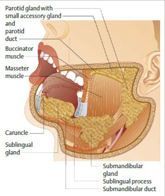
Picture 2.1 Gland major saliva 2
The parotid salivary gland is the largest major salivary gland, weighing about 14-18 grams and measuring approximately 6x4 cm, which is located just anterior and inferior to the auricle. The parotid salivary gland is covered by the parotid fascia which is a continuation of the superficial layer of the deep cervical fascia. The superficial layer of the parotid fascia is continuous with muscular muscular in surrounding, in front to direction masseter muscle, behind towards the sternocleidomastoid muscle and superiorly towards the zygoma. This layer also forms septa into the parotid salivary gland. The deep layer of the parotid fascia separates the parotid salivary gland from the submandibular salivary gland, where this fascia extends towards the mandible anteriorly, towards the styloid process posteriorly, and towards the stylomandibular ligament inferiorly. 1,2
The main blood vessel that supplies the parotid salivary gland is the parotid artery. carotid external. Artery This walk in a way parallel with OS mandibular artery then branches into the maxillary artery and superficial temporal artery. A branch of the superficial temporal artery, namely the transverse facial artery supplies the parotid salivary gland, Stensen's duct and the muscle masseter. 2,3,11
Drainage of the parotid salivary gland is via the posterior facial vein, which is a union of the superficial temporal and maxillary veins. This vein runs beneath the facial nerve and lateral to the carotid artery. It then joins the postauricular vein to form the external jugular vein. facial vein posterior can also join with facial vein front to form facial vein communist, who then flows into the internal jugular vein. 1,3
The parotid salivary glands are closely related to important structures around them, namely the internal jugular vein, the external carotid artery and its branches. lymph glands, branch auriculotemporalis from nerve trigeminal and facial nerves. 1 The facial nerve divides the parotid salivary gland into two parts, namely the majority of the gland is located superficial to the facial nerve, while a small part is located inside and medial to the facial nerve. The facial nerve exits the temporal bone at the stylomastoid foramen, which is located at the junction of the mastoid processes. and the base of the styloid process. When the nerve enters the parotid salivary gland, it divides into two parts, namely the temporo-facial part towards the top and the cervico-facial part towards the bottom. Based on this division, five different areas of the face are innervated by the nerves: (1) Branch temporal that cross arch zygoma going to area temporal; (2) Branches zygoma going to corner lateral eye; (3) Branch open mouth Which going to nose and mouth; (4) Mandibular branch which innervates the muscles of the lower lip and chin; (5) Cervical branch which goes to the neck platysma. 1,3,11

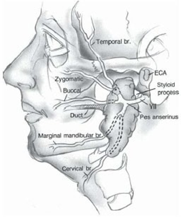
Picture 2.2 . Gland parotid And branching nerve facialist 1
Histologically, the parotid salivary glands are mostly composed of acinar gland cells connected to the salivary duct. These structures are located in a glandular mesenchymal tissue consisting of connective tissue, blood vessels and lymphatics, lymphatic tissue and nerve fibers. 2,3
The duct of the parotid salivary gland (Stensen's duct) is about 6 cm long, begins from the anterior part of the parotid salivary gland and runs horizontally across the masseter muscle approximately 1.5 cm below the zygoma. At the anterior border of the masseter muscle, the duct turns medially and pierces the buccinator muscle through the cheek cavity into the oral cavity through a small papilla opposite the crown of the second maxillary molar tooth. 3,11
The acinar glands are the main producers of saliva, which carry enzymes such as amylase, and sialomucin. The acinar glands contain myoepithelial cells that form a spider-web-like structure and function in the process of emptying saliva production with contractile movements. 3,4 The response of saliva to stimuli depends on the neural reflex carried by system nerve parasympathetic. Nerve parasympathetic gland parotid begins in the inferior salivatory nucleus. Its fibers leave the brain via the glossopharyngeal nerve and through the middle ear, crossing the promontory of Jacobson's nerve. In the tympanic plexus, these nerves enter lesser petrosal nerve, by Because reach the ganglion oticus. Postganglionic fibers from the otic ganglion reach the parotid gland via the temporal auricular part of the fifth nerve. The parasympathetic nerves of the submandibular gland originate from the superior salivatory nucleus. Its fibers enter the nervus intermedius (nerve of Wrisberg) and follow the facial nerve to enter the vertical part of the mastoid. These fibers then leave the nervus seventh in the chorda tympani, through the middle ear, and join the lingual nerve. These fibers follow the lingual nerve to a small ganglion closely associated with the submandibular gland. Postganglionic fibers leave the submandibular ganglion through the substance of the gland. Since severing the chorda tympani nerve and Jacobson's nerve does not always reduce salivary secretion, there must be other parasympathetic nerve pathways that supply the glands. It is thought that these pathways involve the hypoglossal and glossopharyngeal nerves. The sympathetic nerves that supply the major salivary glands arise from the superior cervical ganglion via the arterial plexus. Sympathetic stimulation of the major salivary glands has been reported to cause increased flow followed by a compensatory decrease in flow. Because of the absence of the muscle in glands That Alone, so matter It is believed that this increase in flow may be due to contraction of the myoepithelial , or basket cells associated with the striated ducts.3
The salivary glands constantly produce a certain amount of saliva, even when the body is at rest. In the parotid salivary glands, the amount of secretion when stimulated can reach 4-5 times more than the secretion when resting. 2,4 The total amount of saliva production from all salivary glands is 500-1000 milliliters per day. 2,3 Saliva has several important functions, including lubricating the food bolus and protecting the surface of the oral cavity with a biofilm membrane; maintaining the oral cavity milieu with a pH ranging between; maintaining integrity tooth teeth; effect antimicrobial with content component IgA, IgG, IgM, protein, season, peptide And enzyme; as well as help process tasting and digestion.1,4
2. Tumor Parotid Gland
Salivary gland tumors represent a diverse group of benign and malignant tumors with varying degrees of behavior. The incidence of salivary gland tumors saliva 3%-6% from all over tumor head neck with A little variations over the past few decades. Approximately 70% of salivary gland tumors originate from the parotid gland. Parotid gland tumors, approximately 75% to 80% are benign. 4.5
Table 1 . Distribution neoplasm parotid gland 4
Histological type | % |
Tumor benign |
|
Adenoma pleomorphic | 66.8 |
Warthin's tumor | 22.8 |
Tumor benign other | 10.4 |
Tumor malignant |
|
Mucoepidermoid carcinoma | 32.5 |
Adenoid cystic carcinoma | 15.9 |
Ca ex adenoma pleomorphic | 13.5 |
Acinic cell carcinoma | 10.3 |
Adenocarcinoma NOS | 4.7 |
Basal adenocarcinoma cell | 3.9 |
Clear adenocarcinoma cell | 3.2 |
Myoepithelial carcinoma | 2.4 |
Salivary ductcarcinoma | 2.4 |
Tumor malignant other | 11.2 |
In adults, most salivary gland tumors are benign. Malignant disease accounts for 5% of all head and neck cancers and less from 0.5% from all over cancer, with incidence 2.5-3.0 case per 100,000 per year in the United States. The most common benign tumor of all salivary glands is pleomorphic adenoma, also known as benign mixed tumor . Predisposing factors for neoplastic processes in the major salivary glands include a history of low-dose radiation exposure. 4.5
3 Adenoma pleomorphic
Definition
Pleomorphic adenoma is the most common benign tumor of the salivary glands with an incidence rate of 86%. This type of tumor first was described by Billroth in 1859 and is known by various terms such as mixed tumor, enclavoma, branchioma, endothelioma, enchondroma ), until the term pleomorphic adenoma was introduced by Willis. Its name comes from the pleomorphic architecture that can seen with a light microscope. 6–8 Histologically, pleomorphic adenoma consists of myoepithelial and epithelial cells with different morphological patterns. Adenoma most originate in the superficial lobes but occasionally invade deeper glandular tissue and the parapharyngeal space. Pleomorphic adenomas generally appear as slow-growing, asymptomatic swellings that do not involve the facial nerve. Although most manifest in the parotid glands, pleomorphic adenomas can also be located in the salivary glands of the hard and soft palate, upper lip, cheeks, tongue, and floor of the mouth. 6,8 Pleomorphic adenomas can develop into malignancies, namely into a benign but metastatic subtype, namely metastatic pleomorphic adenoma, or a mixed malignant tumor called carcinoma ex-pleomorphic adenoma and carcinosarcoma. 8 Carcinosarcoma and metastatic pleomorphic adenoma are very rare with metastatic pleomorphic adenoma accounting for 1% of all pleomorphic adenoma malignancies. The World Health Organization (WHO) classifies ex-pleomorphic adenoma tumors that show malignant transformation as epithelial tumors. However, in rare cases, metastases may occur. happen without transformation malignant This, And with thus There is debate regarding the nomenclature and diagnostic criteria of this tumor. This controversy is caused by the histology of cases of benign metastatic pleomorphic adenoma that have malignant-like behavior, which is described by the WHO as “a histologically benign pleomorphic adenoma that cannot be explained by manifestations of local or distant metastases”. The appearance of the term 'benign metastatic pleomorphic adenoma' seems to be contradictory. Metastatic pleomorphic adenoma is actually a currently unrecognized and unclassified malignant neoplasm phenomenon and should therefore be considered and treated as a low-grade malignancy. 12
Etiopathogenesis
Predisposing factors for the occurrence of neoplastic processes in the major salivary glands include a history of low-dose radiation exposure. 5 There are two main theories about how salivary gland tumors can arise, but the consensus believes in the multicellular theory, that each form of tumor originates from a specific differentiated cell origin within the salivary gland unit. Excretory stem cells develop into mucoepidermoid and squamous cell carcinomas, while intercalated stem cells can develop into pleomorphic adenomas, adenoid cystic carcinomas, oncocytomas, adenocarcinomas, and acinic cell carcinomas. Several factors are thought to play a role, such as genetics, ionizing radiation, tobacco use, chemical and viral exposures. 3,13 Radiation exposure has also been associated with parotid carcinoma 15 years after the event. Smoking and drinking alcohol have been associated with head and neck squamous cell carcinoma, and scalp and neck skin malignancies have also been known to metastasize to the parotid glands. Some associate exposure to silica dust, nitroasmine, and other chemicals from work. It is said that smoking and drinking alcohol are not related to salivary gland tumors, except for Warth's tumor. 4 The exact etiology of pleomorphic adenoma itself remains unclear, although Martinelli et al. say existence possibility the incident increase from 15 until
20 years after exposure to therapeutic radiation. Only a few study Which show existence connection between adenoma pleomorphic with simian virus 40. Tobacco use, genetic predisposition, and chemical exposure are also thought to play a role in the etiology of the disease. Molecular and cytogenetic studies have postulated abnormalities of chromosomes 8q12 and 12q15. 6
Epidemiology
Pleomorphic adenoma is a common tumor and constitutes two-thirds of all salivary gland tumors. It mostly occurs in the parotid gland (85%) followed by the minor salivary glands (10%) and the submandibular glands (5%). Adult women in the third to fifth decades are most often affected by pleomorphic adenoma. 7
Knight et al. in 2015 found that the mean age of presentation of pleomorphic adenoma in patients who developed metastatic pleomorphic adenoma was 34.3 years with a range of 9-73 years, and the common decade pleomorphic primary adenoma is decades 2nd, the 3rd, And to-
4. A total of 62.9% presented before the 5th decade. The most common was the 2nd decade with 25.9%. cases. Therefore, younger presentation of pleomorphic adenoma may be a risk factor for developing metastatic pleomorphic adenoma. Three cases of metastases Intracranial pleomorphic adenomas have been reported, interestingly, in two of them, primary pleomorphic adenomas presented at ages 9 and 12 years with findings of metastatic pleomorphic adenomas 3 and 51 years later. 12
Knight et al. also found that 72.8% of cases reported local recurrence before the report of metastatic pleomorphic adenoma with 37% showing multiple local recurrences. Currently there is no way to predict which locally recurrent pleomorphic adenomas have the potential to give rise to metastatic pleomorphic adenomas. Some have suggested that locally recurrent pleomorphic adenomas should be investigated for metastases with Positron Emission Tomography (PET). Because of the long latency between resection of pleomorphic adenoma and the development of metastatic pleomorphic adenoma, it is recommended that patients with incomplete excision, spill surgery or relapse local must done act long-term follow-up for pleomorphic adenoma metastases. Practically this may be too difficult and expensive. 12
Histopathology
Histopathologically, pleomorphic adenoma is described as a benign mixed tumor with mesenchymal and epithelial components of the tumor. 4 These cells are single cells that differentiate into epithelial or myoepithelial cells and not just a multiplication of epithelial and myoepithelial carcinogenic cells simultaneously. The tumor has three components: an epithelial component, a myoepithelial cell component, and a mesenchymal component. The diagnosis is made by identifying all three components. The mesenchymal part shows a variable epithelial pattern in a loose fibrous stroma of myxoid, chondroid, mucoid, fibroid, or osteoid type. Myoepithelial cells are polygonal in shape with pale eosinophilic cytoplasm. 4,6
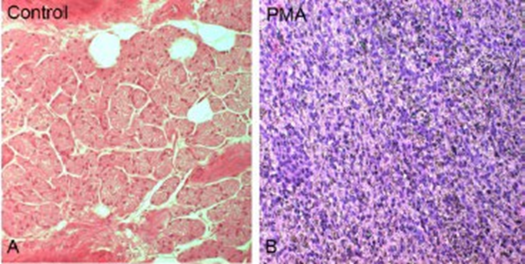
Picture 2.3 Comparison difference structural between control with pleomorphic adenoma of the parotid gland stained with HE. Parotid control network
(A) compared to with pleomorphic adenoma Which arranged on cell epithelium and mesenchyme with different lineage differentiation. 7
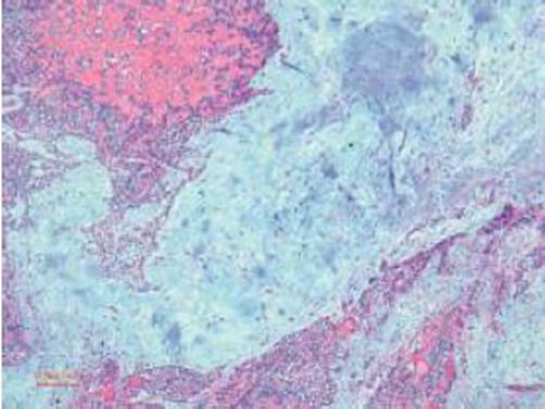
Picture 2.4 Adenoma pleomorphic looks a lot matrix myxochondroid And cell myoepithelial (50× coloring H&E) 6
Specific characteristics of the capsule that may be associated with the likelihood of recurrence, such as the presence of a tumor capsule incomplete, pseudopodia, or satellite nodules (Figure 2.5) . Zbaeren and Stauffer proposed the following nomenclature: complete capsule assumes complete encapsulation of the tumor tissue within an anatomically intact fibrous capsule. Pseudopodium represents a “re-encapsulated” secondary nodule separated only by a layer of capsular fibrous tissue from the main tumor mass but localized within the main tumor capsule. On a sectional plan, pseudopodia may appear as mushroom-like capsular protrusions or as small nodules that are clearly closely associated with the main tumor and separated only by a fibrous capsule, but no normal fat or salivary tissue is visible between them. Final, nodule satellite is nodule tumor different in near primary tumor but outside the tumor capsule main, separated by salivary or fatty tissue without any connection to the main tumor. 11
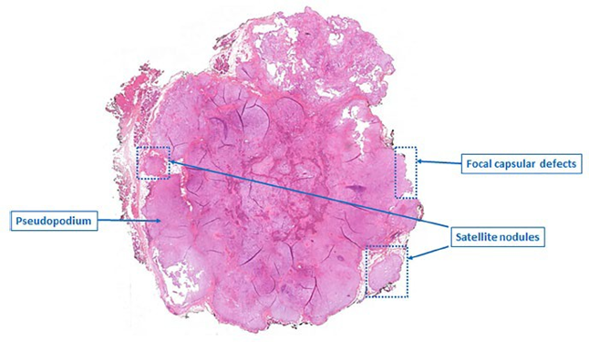
Picture 2.5 Picture adenoma pleomorphic parotid all in all with typical capsule characteristics 11
Symptom And Sign Clinical
The primary site of occurrence is in the parotid gland, which manifests as swelling in the ramus of the mandible in front of the ear and occurs in the superficial lobe. Hard, uneven nodular lesions are the picture. If degeneration cystic shallow And No show fixation, then it can be palpated. In most cases, there are no symptoms, including the absence of pain or facial nerve involvement. 7
In a study conducted by Sungur et al. , asymptomatic parotid mass was the chief complaint in 77.3% of cases. Thirty-two patients (22.7%) showed signs of edema, fluctuation, tenderness, and/or pain. Four cases (1.7%) of facial paralysis were noted. Intraoral involvement was seen in three (1.3%) patients with deep lobe tumors. Symptoms can appear at any age between a few weeks to 12 years. 7
Pleomorphic adenoma can develop into a benign but metastatic subtype. Where this benign tumor most often experiences distant spread to the bones, lungs and lymph nodes. 4
In cases of metastasis to the lymph nodes, the location of the lymph nodes is determined based on the division of the lymph node level. lymph nodes in the neck, as shown in Figure 2.6.
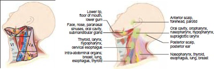
Picture 2.6 Distribution level gland sap clear on neck (A) And regional lymph node groups drain a particular primary site (B). 5
The American Academy of Otolaryngology-Head and Neck Surgery has modified the above system by dividing levels I, II, and V into categories A and B for each level.
Level I
The submental or level I (IA) group includes the lymph nodes between the anterior belly of the digastric muscle from the lower border of the symphysis cephalad of the mandible to the caudal hyoid bone. The submandibular or level I (IB) group includes the lymph nodes in the triangular area bounded by the anterior and posterior belly of the digastric muscle and the inferior border of the body of the mandible. The lymph nodes adjacent to The submandibular salivary glands and those along the facial artery (facial prevascular) are included in this group.
Level II
Level II is the upper jugular group, which includes the lymph nodes around the upper third of the internal jugular vein and the upper part of the spinal accessory nerve, extending from the base of the skull to the bifurcation of the carotid artery or at the level of the ribs. hyoid. The posterior border of this level is the posterior border of the sternocleidomastoid muscle, and the anterior border is the lateral border of the sternohyoid muscle. The lymph nodes anterior to the spinal accessory nerve are called level IIA, and the lymph nodes posterior to it are called level IIB.
Level III
Level III is the midjugular group, which includes the lymph nodes. in about the middle third jugular vein internal of the hyoid bone to the inferior border of the cricoid cartilage. The anterior and posterior borders are the same as those of level II.
Level IV
Level IV is the lower jugular group, which includes the lymph nodes. around the lower third of the internal jugular vein from the inferior border of the cricoid cartilage to the clavicle. The anterior and posterior borders are the same as those of levels II and III.
Level V
Level V is the posterior triangular group, which includes the lymph nodes around the lower part of the spinal accessory nerve and along the transverse cervical veins. It is bounded by triangle formed by the clavicle, the posterior border of the sternocleidomastoid muscle, and the anterior border of the trapezius muscle. Level V is divided into two level by A field as high as limit inferior bone cricoid cartilage. Level VA is superior to this plane, and level VB is superior to this plane. lower than it. In general, the VA level includes the accessory chain lymph nodes and the VB level includes the transverse cervical chain and supraclavicular lymph nodes.
Level VI
Level VI is the central compartment group, which includes the lymph nodes in the prelaryngeal, (Delphian), pretracheal, paratracheal, and tracheoesophageal grooves. The boundaries are cephalad to the hyoid bone to the suprasternal notch caudad and between the medial borders of the carotid sheaths.
Level VII
Level VII is the anterosuperior mediastinal group, which includes the lymph nodes in the anterosuperior mediastinum and the tracheoesophageal groove, extending from the suprasternal notch to the innominate artery.
Diagnosis
Diagnosis made based on taking sample network And radiographic examination.
Inspection Support
Due to its rarity and morphological diversity, its diagnosis is difficult. Imaging plays an important part in the process of staging salivary gland cancer and offers important information for the precise localization of salivary gland tumors (e.g., lobar localization). superficial and deep) and differentiate between benign and malignant. Pleomorphic adenomas seen on USG are generally hypoechoic in texture. They usually have well-defined borders with lobules, either with or without posterior acoustic enhancement. 7
On computed tomography (CT) scan, pleomorphic adenoma usually appears as a globular mass with a homogeneous soft tissue density that is smooth or lobulated. Larger masses may show necrosis. Fewer foci of calcification are common. The method of choice for analyzing salivary gland cancer is magnetic resonance imaging (MRI). MRI is similar to CT scan; smaller masses appear well-circumscribed and homogeneous, whereas larger tumors appear heterogeneous. MRI is preferred because it provides better delineation, intricate tumor margins, and tumor location relative to surrounding tissue. 6,7 In cases of malignant transformation, postcontrast short tau inversion recovery (STIR) images help define perineural tumor spread. MRI is helpful in differentiating recurrence from posttreatment changes. Assessment of skull base intrusion Cortical tumors often involve postcontrast computed tomography (CT) imaging; they show diffuse, patchy radiolucency and depression-like bone resorption. PET-CT is an efficient way to identify distant salivary gland cancer metastases. 7
Fine Needle Aspiration Biopsy
Fine needle aspiration biopsy/ cytology (FNAB) and core needle biopsy techniques are used to obtain tissue samples that can be performed in an outpatient clinic. FNAB is the primary diagnostic tool for lesions. gland parotid. The value increase when reported by expert experienced cytopathologist in the diagnosis of salivary gland disease. Because of the potential for cell spread and subsequent problems, many experts do not recommend incisional biopsy. The preferred method for obtaining histologic samples and evaluating tumor subtype and severity is fine needle aspiration (FNA). Salivary gland tumors can be accurately diagnosed preoperatively using FNAB for make decision therapeutic, Where help separate benign tumors from malignant tumors. 7 High sensitivity and specificity values (92.6% and 98.4%, respectively) of the cytological diagnosis of pleomorphic adenoma were reported by Viguer et al. in 1997. Carrillo et al. obtained similar results (92.6% and 98.4%, respectively ). 97% and 98%). Data from Fikova et al. showed slightly lower values (sensitivity 88.83% and specificity 96.23%), but confirmed that FNAB is a reliable method in the diagnosis of pleomorphic adenoma.
Another preoperative diagnostic that can be done is core needle biopsy . However, this procedure is more extensive, performed under local anesthesia, and the risk of tumor spread is still controversial. 14 Although the tumor is encapsulated, it is still excised with sufficient margins and involves the surrounding normal tissue. This is because the pseudopods show microscopic extension into the surrounding tissue due to dehiscence in the false capsule. Therefore, incisional biopsy is avoided to prevent tumor cell spillage. 6
Management
The treatment of choice for these tumors is excision with an adequate margin of tumor-free tissue. The standard of care for pleomorphic adenoma is complete excision of the tumor via total or superficial parotidectomy. 13 The surgeon must consider the size and position of the tumor, as well as its vascularity, malignancy, and relationship to important structures including the oropharyngeal airway, neck, and vascular bundles, when determining the best surgical approach. Previously, the procedure standard treatment tumor benign gland parotid is enucleationtumor contents, leaving the capsule in place. 7 Enucleation results in a very high recurrence rate. 11
Surgical modalities on the parotid gland are defined as follows: dissection extracapsular considered as appointment tumor with cuff of parotid tissue without the intention of exposing the main trunk or branches of the facial nerve. If the main trunk is intentionally exposed before tumor dissection and only a portion of the superficial lobe is removed along with the tumor, the procedure is defined as a partial superficial parotidectomy. Removal of the entire parotid gland lateral to the facial nerve is defined as a lateral parotidectomy, while extirpation of the entire parenchyma of the gland while preserving the facial nerve is referred to as a complete parotidectomy. 15
A recent meta-analysis showed that as long as the tumor diameter is less than 4 cm and does not affect the facial nerve, extracapsular dissection may be a good alternative for treating unilateral benign parotid tumors in the superficial lobe. Therefore, it is reasonable to conclude that this is a more complicated technique than extracapsular dissection is necessary for tumors larger than 4 cm to prevent long-term damage to one or more branches of the facial nerve. 7
According to a meta-analysis published in 2002, extracapsular dissection is not recommended for tumors that are closely adherent to nerves because it does not allow complete removal, thereby causing disruption to the capsules and pseudopodia. Although The surgeon intends to remove the tumor using parotid tissue during parotidectomy superficial partial, exposure and the rupture tumor is still possible and is an undesirable outcome. During 20 years of follow-up, there is an 8% chance of recurrence after rupture. If the violence affects lobe in, done total parotidectomy; they may grow medially and involve the parapharyngeal space. Since pleomorphic adenomas may arise in small salivary glands, submandibular glands, plates, nasal septum and rarely on channel hearing external And gland lacrimal, Treatment for pleomorphic adenomas of the smaller salivary glands involves wide local excision along with the affected bone or periosteum. 7
Pleomorphic adenoma has a good prognosis with a cure rate of 95%. The function of radiotherapy is still debated. Jackson et al. gave postoperative radiation to patients whose histology showed that the resection margin of the specimen was not clear of the tumor or to patients whose tumor spilled during surgery. According to Douglas et al. , when total extirpation cannot be achieved, when facial nerve sacrifice is necessary, or after several If it recurs, it is recommended to undergo irradiation. 7
Complications
The most common post-parotidectomy problem was partial facial nerve palsy, observed in 27 patients (35%), followed by recurrence (8%), and total facial paralysis in 4% of cases. Several other problems were observed, including bleeding and wound infection, Frey syndrome, and temporary or permanent facial nerve dysfunction. After surgery, sialocele was also reported as one of the complications. 7 The consent given should include temporary and permanent facial nerve dysfunction, numbness in the ear, excessive sweating (Frey syndrome), sialocele, hematoma, and recurrence. Facial nerve dysfunction and Frey syndrome were less common or partial superficial parotidectomy with nerve dissection compared with total or total superficial parotidectomy. 11
In cases where pleomorphic adenoma affects the superficial lobe of the parotid gland, superficial parotidectomy with preservation is performed. nerve face. This No only reduce duration operation but also reduces the risk of damaging the facial nerve because fewer branches are dissected. In their prospective study, Stathopoulos et al. confirmed that transient nerve palsy was lower after partial superficial parotidectomy than after total and superficial parotidectomy. It is known that the risk of nerve injury is also proportional to the length of the dissected nerve. 7Bittar et al. concluded in their study that facial nerve damage was significantly more likely to occur in tumors measuring 3.0 cm in length and/or 2.0 cm in depth. After superficial parotidectomy, there was an increased risk of facial paralysis secondary to surgery for significant recurrent malignancy. While another study conducted by Kalwaniya et al. concluded that patients with tumors measuring >4 cm and >2 cm in depth or who had undergone resection had an increased risk of immediate facial nerve paresis or paralysis. It was found that 11.3% of patients experienced immediate facial nerve paresis or paralysis. after parotidectomy. 7 The surgical department of FKUI/RSCM found complications of paralysis nerve facialist as much as 18% from operation on tumor benign and 25% of malignant tumor operations. Facial nerve paralysis after parotidectomy surgery can be temporary, which is around 9.3% to 64.6% which can heal itself within 6 months, while permanent paralysis is found in around 8% of cases. Frey's syndrome after parotidectomy occurs due to cross-reinnervation of the autonomic pathway to the parotid gland so that parasympathetic fibers stimulated by smell and taste innervate the sweat glands and blood vessels. As a result, sweat occurs And redness in around skin on region parotid moment chewing. 13
Prognosis
Historically, a very high recurrence rate of 20% to 45% has been observed with simple tumor enucleation. Thus, complete surgical resection with adequate margin of normal tissue is recommended. Currently, tumor resection with partial parotidectomy with preservation facial nerve recommended. Procedure This defined as parotidectomy less than the entire lobe or less than the entire facial web branch is dissected. The rate of facial nerve weakness and Frey syndrome was found to be lower with this approach. 4 A high recurrence of pleomorphic adenoma was found with the enucleation surgical technique. The first hypothesis for recurrent pleomorphic adenoma was proposed by Patty on year 1950s take notes lobulation small or Tumor pseudopodia seen microscopically outside the presumed capsule, which occurs during uncontrolled enucleation, may potentially break off and remain within the remaining normal salivary gland tissue. The second hypothesis directly implies “ tumor spillage.” that if the tumor capsule is disrupted, tumor cells may become embedded in the surgical wound and some new tumor may slowly start to grow at the surgical site. Other factors that cause recurrence include incisional biopsy of presumed lymph nodes or cysts, peroral approach to parapharyngeal tumors, and implantation of pleomorphic adenoma cells by core needle biopsy or open biopsy. 16 Pleomorphic adenomas can develop into benign but metastatic subtypes. Where these benign tumors most often experience distant spread to the bones, lungs and lymph nodes. 4 Rarely, pleomorphic adenomas can metastasize and remain benign and appear histologically. The subtypes include metastatic pleomorphic adenoma, malignant mixed tumors called carcinoma ex-pleomorphic adenoma and carcinosarcoma. 8–10
Pleomorphic adenoma of the salivary glands, the most common salivary gland tumor, has a very high probability of developing into carcinoma ex-pleomorphic adenoma, the fifth most common salivary gland carcinoma. Rapid growth in the presence of a known pleomorphic adenoma and/or the development of new pain, facial nerve palsy, skin ulceration or fixation, or lymphadenopathy are additional possible manifestations. In addition, patients may have a history of a previous procedure for pleomorphic adenoma. Only 3% of pleomorphic adenoma of the salivary gland recurred after 12.5 years of follow-up, and of these, 6% appeared to show malignant change, so the risk of this transformation is rare. occurred. The recognition of a pleomorphic adenoma component in malignant salivary gland tumors, which were subsequently classified as carcinoma ex-pleomorphic adenoma, was a major supporter of the concept that pleomorphic adenomas of the salivary glands undergo malignant transformation. The benign pleomorphic adenoma component of the tumor This changed in a way morphological And molecular become component carcinoma fierce. 7
CONCLUSION
Salivary gland tumors represent a diverse group of benign and malignant tumors with level incident tumor gland saliva 3%-6% from all over tumor head and neck. Approximately 70% of salivary gland tumors originate from the parotid gland. Pleomorphic adenoma (a benign mixed tumor) accounts for 75% of parotid gland tumors in adults. Adult women in their third to fifth decades are most commonly affected by pleomorphic adenoma. FNAB is a reliable method for diagnosing pleomorphic adenoma. Complete excision of the tumor is the definitive treatment. Most cases of pleomorphic adenoma metastasis occur with a history of recurrence and generally post action result excision incomplete, the possibility of tumor spill on surgical site, or tumor seeding, which is the implantation of tumor cells through hematologic or lymphatic pathways. Even after removal, long-term follow-up is necessary to check for recurrence.
REFERENCE
Walvekar RR, Loehn BC, Wilson MN. Anatomy and Physiology of the Salivary Glands. In: Bailey's Head & Neck Surgery Otolaryngology. 5th ed. Philadelphia: Lippincott Williams & Wilkins, a Wolters Kluwer business; 2014. p. 691–700.
Probst R, Grevers G, Iro H. The Salivary Glands. In: Basic Otorhinolaryngology: A Step-by-Step Learning Guide. 2nd ed. New York: Georg Thieme Verlag; 2006. p. 131–41.
Adams G. Diseases of the Salivary Glands. In: Boies Fundamentals of Otolaryngology: A Textbook of Ear, Nose, and Throat Diseases. 6th ed. Philadelphia: WB Saunders; 1997. p. 305–7.
Oh YS, Russell MS, Eisele DW. Salivary Gland Neoplasms. In: Bailey's Head & Neck Surgery Otolaryngology. 5th ed. Philadelphia: Lippincott Williams & Wilkins, a Wolters Kluwer business; 2014. p. 1760–85.
Shah J, Singh B, Patel SG, Wong RJ. Jatin Shah's Head and Neck Surgery and Oncology. 5th ed. Elsevier; 2020.
Almeslet AS. Pleomorphic Adenoma: A Systematic Review. Int J Clin Pediatr Dent. 2020;13:284–7.
Kalwaniya DS, Meena R, Kumar D, Tolat A, Arya SV. A Review of the Current Literature on Pleomorphic Adenoma. Cureus. 15:e42311.
Meyer MT, Watermann C, Dreyer T, Ergün S, Karnati S. 2021 Update on Diagnostic Markers and Translocation in Salivary Gland Tumors. Int J Mol Sci. 2021;22:6771.
Chen I h, Tu H y. Pleomorphic adenoma of the parotid gland metastasizing to the cervical lymph node. Otolaryngol--Head Neck Surg Off J Am Acad Otolaryngol-Head Neck Surg. 2000;122:455–7.
Ko S, Park KH, Lee JH, Park KN. A case of initially metastasizing pleomorphic adenoma of parotid gland. Rare Tumors. 2022;14:20363613221130155.
Chan Y, Goddard JC, editors. Salivary Glands Diseases. In: KJ Lee's Essential Otolaryngology Head & Neck Surgery. 11th ed. McGraw-Hill Education; 2016. p. 543–63.
Knight J, Ratnasingham K. Metastasising pleomorphic adenoma: Systematic review. Int J Surg. 2015;19:137–45.
Kertanadi N, Sudipta M, Ardika G. Superficial parotidectomy in parotid pleomorphic adenoma. Medicina. 2014;45.
Fikova A, Novak S, Kalfert D, Kuchar M, Zabrodsky M, Dostalova L, et al. Utility of fine-needle aspiration biopsy (FNAB) in parotid pleomorphic adenoma diagnosis and management. Biomed Pap [Internet]. 2023 [cited 2024 Feb 20]; Available from: https://doi.o rg/10.5507/bp.2023.027
Mantsopoulos K, Thimsen V, Gostian AO, Müller SK, Sievert M, Iro AK, et al. Histopathology of Parotid Pleomorphic Adenomas: A “Pleomorphic Approach” to a Demanding Lesion. The Laryngoscope. 2022;132:73–7.
Witt RL, Eisele DW, Morton RP, Nicolai P, Poorten VV, Zbären P. Etiology and management of recurrent parotid pleomorphic adenoma. The Laryngoscope. 2015;125:888–93.
Nouraei SAR, Ferguson MS, Clarke PM, Sandison A, Sandhu GS, Michaels L, et al. Metastasizing Pleomorphic Salivary Adenoma. Arch Otolaryngol Neck Surg. 2006;132:788–93.
Fonseca D, Arya SS, Kodandapani S, Chandini A, Kurapati S, Rao C, et al. Metastasising Pleomorphic Adenoma: A Rare Entity. Indian J Otolaryngol Head Neck Surg Off Publ Assoc Otolaryngol India. 2022;74:6321–3.
Comments