Neonatal hearing loss On Neonate With Factor Risk of Hyperbilirubinemia
I Made Nudi Arthana
Neonatal hearing loss is one of the health problems that has received more attention in this decade. This is due to the development of management to improve children's functional, intellectual, emotional, and social abilities. Neonatal hearing loss generally has a sensorineural type that is bilateral with a degree of severe to very severe. 1,2
LITERATURE REVIEWS
Anatomy And Physiology Ear Organ
The hearing organ "ear" is a paired organ, located on each side of the head. The human ear has a hearing range of 20-20,000 Hz through delivery air, temporary range This more big For frequency Which much higher if through bone conduction. The first part of the ear deals with the transmission of sound to the cochlea sense organ and then the cochlea is responsible for the transduction of vibrations, which is carried out by fine hair cells. The ear is structurally and functionally divided into three parts which are required For hearing normal: ear outside, ear middle, And inner ear (vestibular labyrinth and cochlea). The structure of the ear is shown in Figure 1.7
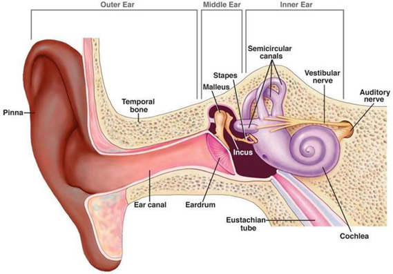
The outer ear consists of the pinna (ear flap) and the external acoustic canal. The pinna protrudes from the side of the skull and is made of cartilage and is completely closed skin. Pinna responsible answer For gather sound vibration And channel it to channel hearing. Pinna very help in localizing voice Because He catch voice, Which more efficient come from front than coming from behind because of the angle. 7
The internal acoustic canal is about 4 cm long and has a hairy outer part and a thinner inner part. The hairy outer part own gland sebaceous And gland sweat , Which both of them together with keratin forming an ear wax . Ear wax growth hair in The outer part of the canal acts as a disinfectant and provides a protective barrier for the ear. 7
The inner ear consists of a membrane tympani, malleus, incus, stapes, muscles tensor timpani And muscle stapedius. Membrane timpani own layer skin outer layer that is continuous with the auditory canal and the inner layer called the endoderm. The outer ectoderm layer is made of stratified squamous epithelium, which shows a unique lateral migration of cells from the center to the edge of the tympanic membrane, where these epidermal cells can be shed. The entire structure of the tympanic membrane is about 0.1 millimeters thick and in the middle ear cavity, membrane timpani cover hole (round) with diameter around 1 cm. Although membrane timpani in a way general called as drum ear, but in a way technical cavity ear middle is drum ear with membrane timpani Which act as skin drum. 7
The three auditory ossicles are the malleus, incus and stapes . These bones conduct sound from the eardrum to the inner ear. The malleus is shaped like a club with its handle embedded in the tympanic membrane, stretch in throughout part middle to on, And his head is at in the cavity ear middle in on membrane tympanic membrane . Which is at in between the other two ossicles, has a thin projection that projects outward called its long process. The stapes is a curved bone consisting of a footplate and an arch. The footplate of this bone is articulated by joints stapedio-vestibular Because cover window oval Which is opening into the vestibular system of the inner ear or cochlea. 7
The human inner ear is located between the middle ear and the acoustic meatus and is called the labyrinth of the ear which can be a bony labyrinth or a membranous labyrinth which is each divided into three parts namely the semicircular canal , vestibule , and cochlea while the membranous labyrinth consists of the semicircular canals, two sac-like structures in the vestibule; namely the saccule And utricle, And channel cochlea. Room between labyrinth membrane And The bones are filled with a watery fluid called perilymph which is obtained from the lymphatic system, and it is similar but not identical to the aqueous humor of the eye and the cerebrospinal fluid. 7 Labyrinth membrane Also own fluid closed Which called endolymph, which has a high potassium concentration, and its composition is different from the perilymph. Endolymph is produced by the vestibular darkcell which has similarities with the stria vascularis, which is part of the cochlea. Endolymph in the membranous labyrinth of the inner ear interacts with hair cells and causes hair cell depolarization by providing a high potassium gradient, resulting in afferent nerve transmission. 7,8
The vestibular system is the sensory system of the inner ear that is important for maintaining postural balance and helping to develop coordination. between position head And movement eye. System This consists of from five organ; three channel half circle Which is at on corner Which appropriate One The same other and control rotation corner (Head) And two organ otolith Which play role important in acceleration linear (movement line straight).
Cochlea is structure bone hollow circular Which coated by epithelial tissue. Although it is bone, it is very important for hearing and transduction. as part from system hearing. Named cochlea from Greek 'kokhliās', meaning snail, because of its coiled shape. The spiral shape cochlea This help him For differentiate frequency Which different, because different but specific spiral areas of the cochlea detect different frequencies. The cochlea consists of three channels lined by epithelial cells filled with fluid. In addition, there is also the organ of Corti, a sensory organ that converts sound energy voice become signal nerve Which delivered through fiber nerve to brain. 7.8
Physiology Hearing
The hearing process begins with the capture of sound energy by the earlobe which then channels it into the external acoustic canal. The external acoustic canal acoustician external act as tube resonance Because amplifies sounds between 3000 and 4000 Hz to increase the ear's sensitivity to those frequencies. The ear responds to very low-intensity sounds because of its sensitivity. The same air pressure on both sides of the tympanic membrane Also allow sensitivity This. Tube Eustachian give the pressure that The same with open in interval short on every swallow the 3rd or 4th. If still open throughout time, somebody even can hear the sound of his own breathing.
The outer and middle ear amplify sound signals because the pinna has a relatively large surface area and channels sound to the tympanic membrane. Which more small Which on its turn own wide surface Which larger than the stapes footplate . This results in hydraulic amplification, that is, smaller movements over a large area are converted into larger movements over a smaller area. The ossicular chain acts as a lever system to amplify the sound. Overall, both the outer and middle ears amplify sound by about 30 dB on its way from the outside to the inner ear. This amplified vibration energy will be transmitted to the stapes which moves it. window oval so that perilymph on scale vestibule move. Vibration forwarded through membrane Reissner Which push endolymph so that it will cause relative movement between the basilar membrane and the tectorial membrane. This process is a mechanical stimulus that causes deflection of the hair cell stereocilia so that the ion channels open and there is a release of electrically charged ions from the cell body. This condition this gives rise to the process depolarization of hair cells so that neurotransmitters are released into the synapse which will cause potential action on the nerves auditory, to be continued to the nucleus auditory to the auditory cortex in the temporal lobe, namely area 39-40. 7,8
Disturbance Hearing On Baby And Child
Definition Disturbance Hearing
Hearing Loss is a term that refers to the partial or complete inability to hear. This can be caused by problems with the ear (outer, middle, and/or inner), the vestibulocochlear nerve (i.e., the eighth cranial nerve or CN VIII), and/or the auditory system. 9 Hearing loss is also defined as any disturbance in the ability to hear sounds at a threshold that is considered normal. 10
Hearing loss is usually categorized as conductive, sensorineural and mixed. Conductive hearing loss results from blockage or disease of the external acoustic canal, tympanic membrane and middle ear, which prevents the transmission of sound energy to the inner ear. Causes of conductive hearing loss range from impacted cerumen and otitis media to fixation of one or more middle ear bones, particularly fixation of the stapes due to otosclerosis. Medical or surgical treatment of most types of conductive hearing loss can often restore full hearing. 11
Sensorineural hearing loss affects the conversion of mechanical sound into neuroelectrical signals in the inner ear or auditory nerve. Mixed hearing loss is the result of damage to the conductive pathways of the ear. part outside and/or middle And nerve or cell hair sensory ear inside . 9,10
Deafness is often used as an alternative to severe hearing loss, when somebody No can hear conversation normal without amplification Hearing impairment. Deafness is a general term for anyone who has a hearing impairment . hearing Which range from light until currently until Enough severe and often benefit from hearing aids. 10
In children, pure tone thresholds averaging more than 15 dB at 500, 1000, 2000, and 4000 Hz are considered outside the normative range, with larger reductions in hearing levels classified by severity. Some levels of severity are Slight hearing threshold 16 to 25 dB; Mild hearing threshold 26 to 40 dB; Moderate hearing threshold 41 to 55 dB; Moderately severe hearing threshold hearing 56 until 70 dB ; Severe : threshold hearing 71 until 90 dB ; profound : hearing threshold more than 90 dB. 10
Factor Reason Disturbance Hearing In Neonates
The most common causes of sensorineural and mixed hearing loss Which nature permanent is congenital cytomegalovirus (CMV; 5% - 20%), abnormality structure bone temporal (30% - 40%), And reason genetics (50%). Many anatomical abnormalities are associated with genetic causes, including branchiootorenal syndrome and CHARGE syndrome. Branchiootorenal syndrome is associated with abnormalities in the second branchial arch derivatives, external ear malformations, hearing loss, and kidney disorders. CHARGE syndrome consists of ( coloboma, heart defects, atresia choanae , growth retardation, genital abnormalities, and ear abnormalities ). Abnormalities ear can in the form of abnormality in the outer, middle or inner ear. 12
Incident disturbance hearing increase along with birth premature and decreases with increasing gestational age and birth weight (1.2% - 7.5% Which born on age 24-31 Sunday And 1.4% - 4.8% on heavy birth weight 750 - 1500 g) and increasing number of comorbidities. NICU-related hearing loss also increases with the combination of hyperbilirubinemia, sepsis, neonatal bacterial meningitis, necrotizing enterocolitis, prolonged ventilation, use of ototoxic drugs. 10 Although congenital CMV is usually the primary cause of hearing loss, it is uncommon; therefore, a genetic or structural etiology is bone temporal other Also can appear as etiology addition. Congenital infections, including syphilis and rubella can cause hearing loss. Rubella, Which in the past is reason most general congenital sensorineural hearing loss, is now rare due to maternal vaccination. 10
Evaluation Disturbance Hearing On Neonate
Assessment of hearing loss in neonates is done by conducting hearing screening on newborns. Hearing screening aims For find disturbance hearing as early as Possible on baby new born so that can to be done immediately habilitation hearing optimal so that negative impact can limited. Technique Which most often used And succeed used on universal newborn hearing screening (UNHS) is: (i) automated auditory brain stem response (AABR) And (ii) otoacoustic emissions (OAE). Technology OAEs and AABRs provide non-invasive recordings of the physiological activity underlying normal hearing function, and both are easy to perform in neonates and infants. 13
OAE recording usually takes <1 minute and can be performed without audiology expertise. The principle of this test is that the sound vibrations emitted from amplifier cochlea normal, flow to hole ear, in where acoustic energy is recorded. During the examination, a small probe is placed in the ear canal, Which to deliver stimulation voice to in system hearing. In a healthy ear, sound stimuli are transmitted through the middle ear to the inner ear, where the outer hair cells of the cochlea produce active responses or emissions. These emissions are captured by a microphone in the probe, analyzed by the filtering unit and a " pass " or "refer" result is automatically displayed on the unit's display. 13
Screening OAE very sensitive (between 85 And 100%) And Enough specific (between 91 and 95%). The main causes of false-positive results in OAE testing are temporary conditions in the external auditory canal (e.g., collapsed ear canal and debris) and middle ear (e.g., presence of amniotic fluid and mucus), as well as high ambient noise levels. However, because OAEs are generated in the cochlea , OAE technology cannot be used to detect dysfunction nerve (nerve VIII And track stem brain hearing), Which can be caused by exposure to ototoxic drugs or hyperbilirubinemia. To diagnose and differentiate these pathologies, the auditory brainstem response is used . 13
AABR plays a very important role in testing the location of the lesion, thus differentiating between conductive hearing loss and cochlear hearing loss . This method is very sensitive to detect pathological mechanisms that cause hearing loss through afferent impulse disturbances, known as auditory neuropathy spectrum disorders. The presence of OAEs and major AABR abnormalities occurring together usually indicates auditory neuropathy. Conversely, the absence of OAEs and normal AABR recordings can improve the diagnosis of hearing loss and usually indicate middle ear problems. 13
Usually, program universal newborn hearing screening (UNHS) is a two-stage approach. The first stage of screening is performed in the first days of life, before discharge from the hospital after birth, and usually involves one or two steps of OAE testing or OAE and AABR in high-risk infants. Children who fail the in-hospital screening test are referred for retesting between 2 and 8 weeks after discharge (second stage) and are tested using OAE followed by AABR. Results stage second Which positive must validated with consultation ENT and audiology, test ABR diagnostic And test electrophysiology other Which done at the age of three months. Finally, all babies who were identified as having hearing loss must get intervention early Which appropriate quick after final diagnosis, and certainly before the age of 6 months. 13
Hyperbilirubinemia
Definition
Hyperbilirubinemia on neonate is a Which general happen. Condition this can known in a way clinical with existence improvement level bilirubin total in the blood > 5mg/dL. 14 This condition will cause symptoms known as Jaundice. Jaundice on baby or jaundice neonatorum is condition clinical on the baby who marked by coloring jaundice on skin And sclera. Hyperbilirubinemia Can caused by physiological or non-physiological processes. Physiologically, bilirubin levels will increase after birth, then settle and then decrease after 7 days of age. However, 3%-5% of neonates who experience hyperbilirubinemia are a pathological process that is at high risk for kernicterus. 15
Physiological Jaundice
Physiological jaundice is the most common clinical manifestation of hyperbilirubinemia in newborns without any health consequences. Serious. Disease yellow Which due to by immaturity physiological which usually appears between 24-72 hours of age and between the 4th and 5th day can be considered as its peak in full-term neonates while in premature infants on the 7th day, disappearing at 10-14 days of life. Unconjugated bilirubin is the most dominant form and usually its serum levels are less from 15 mg/dl. Based on recommendation latest from American Academy of Pediatrics AAP, bilirubin levels up to 17-18 mg/dl are accepted as normal in healthy newborns. 15
Jaundice non- physiological
This type of jaundice is known as pathological jaundice, which is not easily distinguished from physiological jaundice. The appearance of jaundice within 24 hours due to an increase in serum bilirubin exceeding 5 mg/dl/day, a peak level higher than the expected normal range, the presence of clinical jaundice for more than 2 weeks and bilirubin conjugated (dark urine) Which staining clothes) will be categorized under this type of jaundice.
Etiology Hyperbilirubinemia On Neonate
Some etiologies that The underlying cause of hyperbilirubinemia in neonates can be seen from the onset and type of bilirubin that is distributed, namely:
Jaundice beginning
Clinical jaundice in the first 24 hours of life is most likely pathological and is usually the result of isoimmunization (most commonly due to ABO or rhesus incompatibility) or other causes of significant hemolysis. Review of the mother's antenatal records often provides important information. about incompatibility group blood other (for example group (Kell) or the presence of other antibodies. Infection should always be ruled out in a sick newborn with early jaundice. Although jaundice associated with G6PD deficiency appears after 48 O'clock, matter This need become part from investigation on baby with relevant ethnicity (parents of Mediterranean, Asian, or African origin). If hemolysis is excluded, Crigler-Najjar syndrome should be considered. Although rare, syndrome This can cause jaundice not conjugated non hemolytic which worsens rapidly in early life. Infants with G6PD deficiency may also experience non-haemolytic jaundice in the neonatal period. 16
prolonged jaundice
Persistent clinical jaundice in full-term infants at 2 weeks of age and baby premature on age 3 Sunday called as jaundice prolonged. This is a common referral from the public and mostly presents as asymptomatic jaundice. conjugated Which recorded on baby Which breastfed. History giving food, color feces And urine, as well as inspection clinical part big can eliminate pathological causes. 16
The etiology of jaundice due to breastfeeding is still not fully understood . Model on animal show that breast milk can increase bilirubin absorption in channel digest, so that increase circulation enterohepatic And unconjugated bilirubin levels. The higher activity of beta-glucuronidase (which deconjugates intestinal bilirubin) in breast milk compared to formula milk will again increase serum bilirubin levels by increasing enterohepatic circulation. 17
jaundice conjugated
Serum conjugated bilirubin levels >25mmol/liter are generally considered to be conjugated jaundice. Pale chalky stools and dark urine can become symptom related Which important. Inspection beginning must covers the test function heart And inspect abnormality freezing blood. Ultrasonography heart can provide input more carry on in cases Which suspected as obstructive jaundice. Further investigations can rule out congenital infection, sepsis, glucosemia, and aminoacidemia. Premature infants receiving total parenteral nutrition often have an increase in the conjugated bilirubin fraction that resolves gradually after total parenteral nutrition is discontinued. One of the causes of this obstruction is biliary atresia. Biliary atresia is a rare disorder. In 10% to 20% of patients, it is associated with other congenital abnormalities. Its etiology is still largely unknown and the most widely accepted theory is that an unknown exogenous factor triggers a series of inflammatory events that restrict self on individual Which own trend genetics during embryonic or perinatal period. 17Pathophysiology Hyperbilirubinemia
Bilirubin produced from catabolism heme, product breakdown hemoglobin, in the Reticulo Endothelial System (RES). First, heme is converted to biliverdin, releasing iron and carbon monoxide through the action of the enzyme heme oxygenase. Biliverdin is then converted to bilirubin by the enzyme biliverdin reductase. This unconjugated bilirubin is hydrophobic and is transported in the circulation to the liver bound to albumin, where it is conjugated with glucuronic acid in the smooth endoplasmic reticulum by the enzyme uridine diphosphate-glucuronosyltransferase (UGT). Conjugated bilirubin is water-soluble and is then excreted in the bile and into the digestive tract, where where part big excreted in feces after metabolized by intestinal bacterial flora. Some conjugated bilirubin is deconjugated in the gastrointestinal tract by the action of beta-glucuronidase and reabsorbed via the enterohepatic circulation. 18
Newborns have higher total serum bilirubin levels than adults due to higher hemoglobin levels at birth, along with shorter RBC lifespan and limited conjugation ability. on heart baby Which new born. By Because That, baby Which new born Term infants typically have peak serum bilirubin concentrations of 5 to 6 mg/dL compared with adult levels of <1 mg/dL. Pathological jaundice in neonates is related to increased bilirubin production in the RES, impaired hepatic absorption, inadequate conjugation of bilirubin, and/or increased enterohepatic circulation of bilirubin. 18
In severe hyperbilirubinemia, unbound and unconjugated bilirubin across blood brain barrier And bound with stem brain, hippocampus, brain small, globe pallidus, And core subthalamic. At the cellular level, bilirubin inhibits certain mitochondrial enzymes, inhibiting synthesis DNA And protein, induce termination strands DNA, and inhibits phosphorylation. Bilirubin also interferes with tyrosine uptake and alters the normal function of the N-methyl-D-aspartate receptor ion channel. These mechanisms are involved in the pathogenesis of bilirubin toxicity that manifests clinically as bilirubin-induced neurological dysfunction and bilirubin encephalopathy. The duration of bilirubin exposure and the amount of bilirubin in the brain determine the severity of brain damage. However, total serum bilirubin levels do not correlate well with bilirubin toxicity in the absence of hemolysis. Premature infants are even more susceptible to the toxic effects of free unconjugated bilirubin. This is partly related to the relatively lower serum albumin levels, CNS immaturity, and concomitant comorbidities such as intraventricular hemorrhage, periventricular leukomalacia, sepsis, necrotizing enterocolitis, and bronchopulmonary dysplasia. 18 Hyperbilirubinemia conjugated caused by by abnormality on absorption, metabolism, transportation, and/or excretion salt bile And bilirubin. This disorder increase sour bile in heart Which push proliferation channel bile and fibrosis. Sour bile Also responsible answer on inflammation And hepatocyte apoptosis leading to hepatocellular injury and cirrhosis. Decreased bile secretion in cholestasis causes malabsorption of fat and fat-soluble vitamins which often leads to failure to thrive with deficiencies of vitamins A, D, E, and K. 18
Influence Hyperbilirubinemia to Disturbance Hearing
The main concern with excessive hyperbilirubinemia is the potential effect neurotoxic, although can Also happen lesion on cells others. This remains a significant problem despite advances. in maintenance neonate jaundice (hyperbilirubinemia). Literature Others explain that severe hyperbilirubinemia that is not treated during the neonatal period will cause high bilirubin levels and be toxic to development. baby. On baby aterm, symptom hyperbilirubinemia is child weak and lazy drink Which will to be continued become choreoathetoid cerebral palsy, retardation mentally And deaf sensorineural. 19,20
There is evidence that even moderate elevations in bilirubin levels can increase the risk of infants developing cognitive disorders, perception, motor And auditory. Researches prospective controlled studies have revealed neurological and cognitive impairment in children who had elevated indirect bilirubin levels in infancy. Extensive statistical studies of healthy term infants, such as those reported by The National Collaborative Perinatal Project, have detected an association between low levels of hyperbilirubinemia that are usually untreated with sequel neurological And motor light. Researches clinical and pathological Which more new Again has prove that level bilirubin What was previously considered safe turned out to be dangerous. 19,20
Recent literature suggests that moderate hyperbilirubinemia in healthy term neonates may be unsafe for their brains. Bilirubin can enter to brain when No bound with albumin or No conjugated or free (Bf) or if there is damage to the blood-brain barrier. Bilirubin is formed from hemoglobin (picture 2), around 75% of it from hemolysis And 25% from erythropoiesis that is not effective. Hemoglobin is first converted to biliverdin through an adenosine triphosphatase-dependent reaction catalyzed by heme oxygenase, producing a molecule of carbon dioxide for each molecule of biliverdin and ultimately bilirubin. 19,20
Non-toxic biliverdin is catalyzed by biliverdin reductase to unconjugated bilirubin, a natural antioxidant at low levels, but neurotoxic. on level tall. Bilirubin No conjugated nature nonpolar, insoluble in water and bound to serum albumin, so there is only a small amount of unbound bilirubin. conjugated in form No bound or bilirubin No conjugated (Bf), but precisely Bf this is it Which free enter in brain, fluid interstitial And cerebrospinal fluid, And responsible on its neurotoxicity. Bf easy cross the barrier blood brain, However bilirubin Which bound on albumin No can enters the brain unless there is a disruption of the blood-brain barrier, then staining will result yellow Which wide. On condition toxic, bilirubin No conjugated who does not bound or Bf-lah (bilirubin indirect) Which There is in brain, No bilirubin Which bound on albumin. 19,20
Bilirubin affects mitochondrial function by inhibiting mitochondrial enzymes, interfering with DNA synthesis, inducing DNA breakdown, inhibiting protein synthesis, disrupting oxidative phosphorylation and inhibiting tyrosine uptake (a marker for synaptic transmission). Bilirubin has an affinity for phospholipids forming precipitates that adhere to brain cell membranes. The mechanism of bilirubin toxicity has been inferred from studies using pathophysiologically relevant bilirubin concentrations, namely the levels of unconjugated bilirubin expected to be found in the central nervous system of infants with hyperbilirubinemia. 19,20
Several studies have been reported to prove that the toxicity model neuronal selective happen resemble incident ischemia brain. Ion homeostasis calcium (Ca+) is mechanism base main Which cause death neuron cell And improvement excitability neurons. Lots neuron use proteins as calcium ion buffers to maintain low intracellular calcium levels. Experiments on icteric mice showed a delay in calcium ion activity and (CaM kinase II), a substance required by cell protein kinase in the phosphorylation process. In vitro it was found that bilirubin hinder activity CaM kinase II, Which considered relate with various important neuron functions, such as: neurotransmitter release, changes in ion conductance regulated by calcium and neuroskeletal dynamics. 19,20
In the brain, susceptibility to the neurotoxic effects of bilirubin varies according to cell type, brain maturity and brain metabolism. Conditions that affect the blood-brain barrier such as: infection/sepsis, acidosis, hypoxia, hypoglycemia, head trauma and prematurity can affect the entry of bilirubin into the brain. 19,20
Unconjugated bilirubin that enters the brain is mainly in free form or anionic bilirubin, bound to phospholipids and gangliosides on the surface of the neuron plasma membrane. The bond between the anionic bilirubin-phospholipid complex is an unstable bond. Anionic bilirubin takes hydrogen ions and forms bilirubin acid which adheres firmly to the membrane. Acid bilirubin the will cause damage on membrane plasma so that can cause bilirubin anion enter to in cell neurons. Bilirubin anions that enter the cells will bind to phospholipids in the membranes of subcellular organelles such as mitochondria and the reticulum. endoplasm and nucleus. This bond will cause the formation of bilirubin acid and membrane damage at the subcellular level. This damage has an impact on multisystem enzymes and causes damage to neuron cells. 19,20
One form of bilirubin neurotoxicity is an abnormality of the auditory system in hyperbilirubinemia. Based on evidence from audiometric tests, bilateral dominant hearing loss is found at high frequencies and symmetrical with abnormal voice development function. This is related to lesi pathological on nucleus cochlear. Bilirubin Which there is on brain can damage the central auditory nuclei and vestibular pathways, cerebellar nuclei and basal ganglia which are associated with vestibular hyperreactivity. 19,20
DISCUSSION
Hyperbilirubinemia in neonates is a common condition. Condition This can be known in a way clinical with existence improvement total bilirubin level in the blood > 5mg/dL. This situation is caused by a combination of increases catabolism heme And immaturity physiological liver in conjugation and bilirubin excretion. Neonatal jaundice occurs in more than 80% of newborns. Although mild jaundice is physiologic and may be neuroprotective, severe hyperbilirubinemia can lead to neurologic dysfunction and death. 15,21
Clinically, neonatal jaundice shows symptoms such as yellow discoloration of the skin, sclera and nails. These symptoms are known to appear when the levels of bilirubin total blood 5-7 mg/dl. Matter This influenced by improvement unconjugated (indirect) bilirubin which is the most elevated form often occurs in hyperbilirubinemia. 21
Kernicterus is a bilirubin-induced brain damage that is most commonly seen in infants. The risk of developing kernicterus is significantly increased in infants with bilirubin levels >25 mg/dL, while levels >30 mg/dL are associated with very high risk and irreversible damage. Any event that causes increased bilirubin production or decreased elimination can lead to hyperbilirubinemia and thus kernicterus. This includes, but is not limited to, polycythemia, hemolysis consequence isoimmunization Rh And disabled default from enzyme Which involved in bilirubin metabolism. 22
The parts of the brain most frequently affected are the basal ganglia, hippocampus, body geniculate And core nerve cranial. Bilirubin Which passing through the blood brain barrier will be deposited in the basal ganglia of neonates, often in the globus pallidus and subthalamic nucleus sectors H2 and H3 in the hippocampus, substantia nigra, reticular formation of the pons, inferior olivary nucleus, and cranial nerves such as nerve oculomotor, facial, vestibular And nucleus ventrocochlear, Which may cause neurologic sequelae, including sensorineural (retrocochlear) hearing loss.2 In A study by The Boss on year 2018, find there is an increased risk factor of neonatal hyperbilirubinemia for hearing loss. In a case-control study, 200 full-term infants with bilirubin levels higher than 20 mg/dl who were admitted to the neonatal intensive care unit at Ghaem Hospital, Mashhad during 2 years. Profile of infants with disturbance hearing (n = 60) compared to with baby new born jaundice with normal hearing (140 newborns) as a control group. ABR test was used to assess the hearing status of infants after discharge from the hospital. It was found that sensorineural hearing loss in infants with severe hyperbilirubinemia was found to be 4.8%. The risk of hearing loss in jaundice infants with bilirubin levels above 20 mg/dl is 10-50 times higher. 2
Similar results were also found in a study by Ahmed and colleagues in 2022 who conducted a prospective observational study conducted on 234 patients during the period November 2019 to April 2021 on all patients treated for hyperbilirubinemia in ENT and Pediatrics department at the hospital education tertiary. From 234 neonate with hyperbilirubinemia, 13 (5.55%) had hearing loss. 57 (24.35%) of 234 neonates showed hearing loss. hearing on OAE on moment included in study. From 57 the, 48 (20.5%) own comorbidity addition like birth premature on 30 (12.82%), heavy body born low on 8 (3.41%), syndrome
meconium aspiration in 11 (4.70%), sepsis in 3 (1.28%) experienced hearing loss And 13 (5.55%) experience disturbance hearing Which settle down at follow-up at 6 months and required further management.
Significant association was noted with cases of hyperbilirubinemia in preterm neonates and neonates with sepsis. A total of 97 (41.45%) preterm neonates suffered from hyperbilirubinemia. Of the 97, 30 (12.82%) had AED referral at the time of enrollment and 8 (3.41%) had brain stem evoked response audiometry. test (BERA) Which No normal on age 6 month. Mark p <0.0001 and therefore statistically significant.
In addition to increased serum bilirubin levels, the length of exposure to bilirubin in infants is also a risk factor for hearing loss in infants. Research by Beseli on year 2020 find existence correlation on long exposure to disturbance hearing. Study This done in a way prospective study on 41 full-term newborns hospitalized for indirect hyperbilirubinemia. Results showed that neurodevelopmental disorders were found. at 5 (12.2%) patient. Hearing loss was found in 4 (9.8%) patients. Two of these patients had auditory neuropathy spectrum disorders and two other patients had cochlear hearing loss. The total serum bilirubin levels of all these patients were above 25 mg/dl. The exposure time to sTB levels above 20 mg/dl was significantly longer in patients with neurological dysfunction and pathological ABR results. This study suggests that kernicterus can occur in term newborns with severe hyperbilirubinemia (sTB>25 mg/dl) without significant hemolysis. Not only the bilirubin levels, but also the duration of exposure to high bilirubin levels may be effective in the development of bilirubin neurotoxicity. 25
Study other also looking for predictors of disruption hearing on unconjugated bilirubin levels compared to total bilirubin as. Nam and colleagues in 2019 conducted a study on 70 premature infants with hyperbilirubinemia who failed universal newborn hearing screening with automated ABR. Diagnostic ABR was performed within 3 months of birth. Results Which found Concentration UCB beginning And maximum which is measured For baby premature group ABR diagnostic ≥40 dB nHL (n=30) was statistically higher compared to the ABR ≤35 dB nHL group (n=40) (P=0.031 and P=0.003, respectively). On follow-up ABR examination, 13 of the ABR ≥40 dB nHL group showed complete recovery, but 17 had no change or worsened. From these results, it is concluded that unconjugated bilirubin (UCB) is a better predictor of bilirubin-induced hearing loss than serum total bilirubin in premature infants evaluated by serial ABR. Serial ABR testing may be a useful non-invasive method for evaluating hearing loss. hearing consequence bilirubin Which can reversed in a way early on premature baby . 26
CONCLUSION
Hyperbilirubinemia in neonates is a risk factor for the occurrence of disorders hearing sensorineural. Number incident disturbance hearing in neonates with hyperbilirubinemia is still quite high, so it is necessary to routinely conduct hearing screening in newborns. Early detection and treatment Which fast And appropriate on disturbance hearing sensorineural The effects of hyperbilirubinemia will help improve the quality of life of children in the future.
REFERENCE
Nuseir A, Zaitoun M, Albalas H, Douglas M, Canaan Y, AlOmari A, Alzoubi F.Neonatal Screening for Congenital Hearing Loss in the North of Jordan; Findings and Implications. Int J Prev Med. 2021 Dec 1;12:162. doi: 10.4103/ijpvm.IJPVM_383_20. PMID: 35070195; PMCID: PMC8724675.
Boskabadi H, Zakerihamidi M, Moradi A, Bakhshaee M. Risk Factors for Sensorineural Hearing Loss in Neonatal Hyperbilirubinemia. Iranian Journal of Otorhinolaryngology. 2018;30:195-202
Hardani AK, Goodarzi E, Delphi M, Badfar G. Prevalence and Risk Factors for Hearing Loss in Neonates Admitted to the Neonatal Intensive Care Unit: A Hospital Study. Cureus. 2020 Oct 27;12(10):e11207. doi: 10.7759/cureus.11207. PMID: 33269138; PMCID: PMC7704024.
Verstappen G, Foulon I, Van den Houte K, Heuninck E, Van Overmeire B, Gordts F, Topsakal V. Analysis of congenital hearing loss after neonatal hearing screening. Front Pediatr. 2023 May 15;11:1153123. doi: 10.3389/fped.2023.1153123. PMID: 37255573; PMCID: PMC10226668.
Wroblewska-Seniuk K, Greczka G, Dabrowski P, Szyfter W, Mazela J. The results of newborn hearing screening by means of transient otoacoustic emissions-has anything changed over 10 years? Int J Pediatr Otorhinolaryngol. 2017;96:4–10
Boskabadi H, Zakerihamidi M, Moradi A, Bakhshaee M. Risk Factors for Sensorineural Hearing Loss in Neonatal Hyperbilirubinemia. Iran J Otorhinolaryngol. 2018 Jul;30(99):195-202. PMID: 30083525; PMCID: PMC6064763.
Sheikh A, Bint-e-Zainab, Shabbir K, Imtiaz A. Structure and Physiology of the Human Ear Involved in Hearing [Internet]. Auditory System - Function and Disorders. IntechOpen; 2022. Available from: http://dx.doi.org/10.5772/intechopen.105466
Pulkki V, Karjalainen M. Physiology and anatomy of hearing. Communication Acoustics. 2015 Jan 16;111–32. doi:10.1002/9781119825449.ch7
Michels TC, Duffy MT, Rogers DJ. Hearing Loss in Adults: Differential Diagnosis and Treatment. Am Fam Physician. 2019 Jul 15;100(2):98-108. PMID: 31305044.
Lieu JEC, Kenna M, Anne S, Davidson L. Hearing Loss in Children: A Review. JAMA. 2020 Dec 1;324(21):2195-2205. him:
Cunningham LL, Tucci DL. Hearing Loss in Adults. N English J Med. 2017 Dec 21;377(25):2465-2473. doi: 10.1056/NEJMra1616601. PMID: 29262274; PMCID: PMC6457651.
Neumann, Katrin, et al. "Newborn and infant hearing screening facing globally growing numbers of people suffering from disabling hearing loss." International Journal of Neonatal Screening 5.1 (2019): 7.
Wroblewska-Senju, K., Dabrowski, P., Szyfter, W. et a. Universal newborn hearing screening: methods and results, obstacles, and benefits. Pediatr Res 81, 415–422 (2017). https://doi.org/10.1038/pr.2016.250
Rohiswatmo R, Amandito R. Hyperbilirubinemia On neonate >35 week in indonesia; Current Examination and Management. Sari Pediatri. 2018 Oct 19;20(2):115. doi:10.14238/sp20.2.2018.115-22
Ullah S, Rahman K, Hedayati M. Hyperbilirubinemia in Neonates: Types, Causes, Clinical Examinations, Preventive Measures and Treatments: A Narrative Review Article. Iran J Public Health. 2016 May;45(5):558-68. PMID: 27398328; PMCID: PMC4935699.
Mitra S, Rennie J. Neonatal jaundice: aetiology, diagnosis and treatment. Br J Hosp Med (Lond). 2017 Dec 2;78(12):699-704. doi: 10.12968/hmed.2017.78.12.699. PMID: 29240507.
Chee Y Y, Chung PH, Man RM, Man KK. Jaundice in infant and children: causes, diagnosis, and management. Hong Kong Med J. 2018 Jun;24(3):285-
Chen HL, Wu SH, Hsu SH, Liou BY, Chen HL, Chang MH. Jaundice revisited: recent advances in the diagnosis and treatment of inherited cholestatic liver diseases. J Biomed Sci. 2018 Oct 26;25(1):75. doi: 10.1186/s12929-018-0475-8. PMID: 30367658; PMCID: PMC6203212. 10.1001/jama.2020.17647. PMID: 33258894. 292. him: 10.12809/hkmj187245. Epub 2018 May 21. PMID: 29807950.
Old C, Oghalai JS. Audiologic impairment associated with bilirubin-induced neurologic damage. Semin Fetal Neonatal Med. 2015;20:42–46.
Panahi R, Jafari Z, Sheibanizade A, Salehi, Esteghamati A, Hasani S. The Relationship between the Behavioral Hearing Thresholds and Maximum Bilirubin Levels at Birth in Children with a History of Neonatal Hyperbilirubinemia. Iranian Journal of Otorhinolaryngology.2013;25:127- 134
Hegyi T, Kleinfeld A. Neonatal hyperbilirubinemia and the role of unbound bilirubin. J Matern Fetal Neonatal Med. 2022 Dec;35(25):9201-9207. doi: 10.1080/14767058.2021.2021177. Epub 2021 Dec 26. PMID: 34957902.
Hamza A. Kernicterus. Autops Case Rep. 2019 Jan 14;9(1):e2018057. doi: 10.4322/acr.2018.057. PMID: 30863731; PMCID: PMC6394357.
Teixeira MH, Borges VMS, Riesgo RDS, Sleifer P. Hyperbilirubinemia impact on newborn hearing: a literature review. Rev Associate Med Bra (1992). 2020 Jul;66(7):1002-1008. him: 10.1590/1806-9282.66.7.1002. Epub 2020
Ahmed K, Havle A, Sravya YL, Ahmeds S. Assessment of hearing in newborns with hyperbilirubinemia using otoacoustic emissions and brainstem evoked response audiometry. ijhs [Internet]. 2022 Apr. 25 [cited 2024 Jan. 24];6(S3):3564-73. Available from: https://sciencescholar.us/journal/index.php/ijhs/article/view/6524
Besli GE, Metin F, Aksit MA, Saltik S. Long-term Effects of Indirect Hyperbilirubinemia on Auditory and Neurological Functions in Term Newborns. Medeni Med J. 2020;35(1):29-39. doi: 10.5222/MMJ.2020.26986. Epub 2020 Feb 28. PMID: 32733747; PMCID: PMC7384494.
Nam GS, Kwak SH, Bae SH, Kim SH, Jung J, Choi JY. Hyperbilirubinemia and Follow-up Auditory Brainstem Responses in Preterm Infants. Clin Exp Otorhinolaryngol. 2019 May;12(2):163-168. doi: 10.21053/ceo.2018.00899. Epub 2018 Nov 9. PMID: 30404412; PMCID: PMC6453789.Aug 24. PMID: 32844928.
Comments