Mallory Weiss Syndrome
I Made Nudi Arthana
Mallory Weiss Syndrome (SMW) is a condition Which marked as is laceration mucosa superficial longitudinal (tear Mallory Weiss ) in upper gastrointestinal tract, especially at the gastroesophageal junction. Symptoms of SMW including painful stomach, hematemesis, and vomiting heavy. Blood often lumpy and coffee-ground-like, and the stool may be dark like tar (melena). In the United States, SMW accounts for one to 15% of upper gastrointestinal bleeding in adults and less than 5% in children. 1 According to a systematic review and meta-analysis, 37% of studies on SMW were from Asia. However, the review did not provide specific data on the incidence or prevalence of SMW in Asia, including Indonesia. 2
LITERATURE REVIEW
Anatomy
The esophagus is a tube-shaped organ that extends from the connecting digestive system pharynx with stomach. The esophagus is passed through food to reach the stomach in the further digestion process. The esophagus follows a path that is behind the trachea and heart, in front of the spine, and through diaphragm before enter stomach. Esophagus divided become three anatomical segments, namely cervical, thoracic, and abdominal. The cervical segment starts from the cricopharyngeus and ends at the suprasternal notch . This segment is located directly behind trachea, Which relate with esophagus through network tie loose. In part posterior, fascia prevertebral connect esophagus with sixth to eighth cervical vertebrae. The thoracic duct can be found on the left side of the sixth cervical vertebra. The carotid sheath and the lower pole of the lateral thyroid gland can be found on the lateral side of the esophagus in the lower part of the neck. 3,4
The thoracic segment is located between the spine and the trachea in the superior mediastinum, extending from the suprasternal notch to the diaphragm. When the esophagus is followed to direction distal, esophagus passing by in behind arch aorta on level intervertebral discs T4 to T5 and enters the posterior mediastinum. The abdominal segment, the esophagus runs from the diaphragm to the fundus of the stomach. This segment descends through the right diaphragmatic fissure at the level of the 10th thoracic vertebra and enters the gastric cardia at the level of the 11th thoracic vertebra. 3,4
Esophagus generally own long around nine until ten inches (23 until 25 cm) on person mature, with sphincter Which located in every its proximal and distal ends, a lumen lined with mucosa and connective tissue, and an outer smooth muscle composition. The sphincter located in the front, the upper esophageal sphincter, allows food to pass in only one direction into the esophagus, and in the front, the lower esophageal sphincter allows food to pass in only one direction into the stomach. 3,4
Esophagus cervical And sphincter esophagus part on supplied through branches of the inferior thyroid artery. The thoracic esophagus is supplied by the aortic esophageal artery which is located in branch terminal artery bronchial. Segmen abdominal And sphincter esophagus The lower part is supplied by the left gastric artery and branches of the left phrenic artery. These arteries flow to submucosa esophagus as network congested. Blood vein then drains into the superior vena cava from the submucosal plexus. The azygous system provides drainage for the proximal and distal segments of the esophagus while the middle esophagus accept drainage through vessels blood addition from the left gastric vein which branches from the portal vein. 3,4
The lymphatic ducts and lymph nodes together supply the esophagus with drainage. lymphatic. Channels started as endothelium or as sac-like protrusions that are also endothelial. They then converge into larger lymphatic channels that run the length of the esophagus (parallel to the transverse plane). The direction of flow is determined by paired semilunar valves within these channels. These channels then join at different areas to enter their respective regional lymph nodes. Drainage occurs in three parts of the esophagus, with significant connections between each segment. Drainage into the thoracic duct from the deep cervical lymph nodes is achieved via drainage from segment First proximal esophagus. Drainage to node The superior and posterior mediastinum are reached via the lymphatic vessels of the middle third of the esophagus. Finally, the lymphatic vessels from the third most distal segment of the esophagus eventually drain into the gastric and celiac lymph nodes. 3,4
The innervation of the esophagus involves the sympathetic and parasympathetic nervous systems, with primary innervation originating from the vagus nerve and spinal nerves (from segments T1 to T10) via the thoracic and cervical sympathetic trunks. The vagus nerve is responsible for the parasympathetic motor function of the muscles and glands of the esophagus. Nerve chain thoracic And cervical especially is innervation sympathetic and functions to help narrow blood vessels, contract the esophageal sphincter, relaxation wall muscle, together with improvement activity gland and peristalsis. 3,4
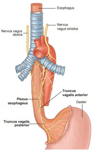
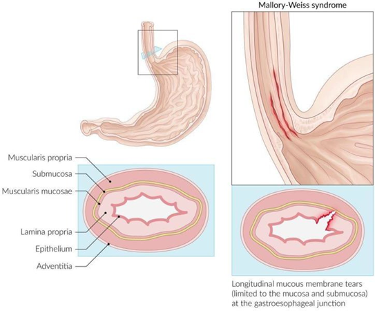
Picture 1. Anatomy Esophagus Normal compared to with SMW. 3.4
Mallory-Weiss syndrome (MWS) is one of the common causes of acute upper gastrointestinal bleeding and is characterized by the presence of superficial longitudinal mucosal lacerations (Mallory-Weiss tears). These tears occur primarily at the gastroesophageal junction and can extend proximally to involve the lower to mid esophagus or distally to involve the proximal part of the stomach ( Figure 1 ). 1 The position of the tear is important to note because it is used to differentiate another clinical condition, namely Boerhaave syndrome. In Boerhaave syndrome, the entire thickness of the esophageal wall is torn or can be called a transmural tear ( Figure 2 ). 3,4 In contrast, the tear that occurs in MWS is a non-transmural tear.
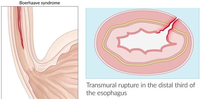
Picture 2. Anatomy Esophagus on syndrome Boerhaave.3.4
Physiology
The main function of the esophagus is to secrete mucus and transport food that enters the mouth through the throat and into the stomach. The upper esophageal sphincter (pharyngoesophageal sphincter) is a circular band of muscle tissue that normally remains closed in position contract. Moment swallow, muscles the relax in a way while and allow material or bolus in form food, drink, mucus, and water saliva from laryngopharynx enter to in esophagus. Furthermore, bolus move into the body of the esophagus. Peristaltic movements propel the bolus down the esophagus through primary and secondary peristalsis. During the pharyngeal stage of swallowing, the muscular walls of the pharynx contract providing a powerful initial peristaltic movement. from bolus And send bolus through sphincter pharyngoesophagus with kinetic energy. This peristaltic wave continues into the esophagus and is primary peristalsis. If primary peristalsis is insufficient it is recognized as continued distension esophagus after peristaltic primary, peristaltic secondary initiated and continues until the bolus is successfully moved to the stomach. 3,4
The lower esophageal sphincter (cardiac sphincter and cardioesophageal sphincter) is located slightly more than one inch (about 3 cm) near the junction of the esophagus and stomach. Similar to the upper esophageal sphincter, this sphincter is normally contracted and closed, primarily preventing gastric contents from entering the esophagus. This sphincter is controlled involuntarily and is triggered to open during esophageal peristalsis, thereby allowing the bolus to be propelled into the stomach and completing the primary function of the esophagus. 3,4 However, the material that distributed by esophagus going to stomach Also can issued from body from the stomach And go out from mouth in case vomit, eructation , And on moment gag reflex is initiated. However, this function is usually undesirable because it can cause malnutrition and damage may occur to the esophagus if it occurs continuously. 3,4
Risk Factors
Mallory Weiss Syndrome (SMW) own a number of factor risk, including consumption alcohol heavy Which is factor most important Because around 50%
Up to 70% of patients diagnosed with SMW have a history of heavy alcohol consumption. The severity of upper gastrointestinal bleeding with SMW has also been reported to be higher in the presence of portal hypertension and varices. esophagus. Besides That, connection between hernia hiatus (protrusi organ, usually the upper part of the stomach into the chest cavity through the esophageal opening of the diaphragm) and SMW is still a matter of debate. Hiatal hernia was found in a number of cases of SMW, while a case-control study found no difference incident hernia hiatus between patient with SMW And group control. 1 Another fairly frequent risk factor is hiccups. Previous studies have found that patients who visit hospitals with complications and symptoms of MWS experience persistent hiccups. A meta-analysis also revealed that hiccups are associated with the development of MWS. 2
Other risk factors include bulimia nervosa, hyperemesis gravidarum, and gastroesophageal reflux disease (GERD). All of these conditions involve regurgitation of gastric contents into the esophagus. However, in a significant number of patients (approximately 25% of cases), no such risk factors are identified. SMW can also be triggered by repeated acts of sudden increase in intra-abdominal pressure such as retching , vomiting, straining, coughing, cardiopulmonary resuscitation (CPR), or blunt abdominal trauma. 1,2 Meanwhile, iatrogenic SMW Also Possible happened even though seldom. This condition can happen as a complication of invasive procedures such as upper gastrointestinal endoscopy or echocardiography. trans-esophageal (TEE). Procedure endoscopy channel digest part The upper arm itself has a complication rate of only 0.07% to 0.49% for developing SMW, so the risk of occurrence is low. 1,2,5
Diagnosis
The exact mechanism of Mallory-Weiss syndrome (MWS) is unknown, but it has been suggested that when intra-abdominal pressure suddenly and significantly increases, gastric contents with that pressure will flow proximally into the esophagus. This excessive pressure of gastric contents Which cause tear mucosa longitudinal Which can reach arteries and vein submucosa, so that cause bleeding channel digest part on
(SCBA). 6,7 SMW diagnosis can be established through anamnesis, physical examination and supporting examinations as follows.
Anamnesis
Mallory-Weiss syndrome may be asymptomatic in mild cases. In 85% case, symptom Which appear is hematemesis, start from blood-tinged mucus, massive bright red to dark bleeding. 1,6,8 If the blood is fresh, the color will be bright red. Patients with SMW will generally experience vomit that is dark in color, lumpy, and resembles coffee grounds. Patients with SMW usually experience violent vomiting followed by episodes of hematemesis. Hematemesis can be an insignificant amount, for example one spoonful Eat, but Also can amount to significant until showing symptoms of hemodynamic instability. Abdominal or chest pain may also be complained of in SMW associated with severe vomiting. 6,7
If happen bleeding massive, symptom other like melena, Dizzy, or syncope can appear although seldom, especially on patient with history of excessive alcohol consumption. 1,2,8 Melena is a darker stool caused by the presence of partially digested blood originating from a Mallory-Weiss tear. When the tear bleeds, blood enters the stomach, undergoing decomposition, And consequence decomposition This, give color black tar-like stool. Melena is a typical symptom of SCBA bleeding such as bleeding in the esophagus that occurs in SMW. 6,7
Weakness can be a symptom associated with anemia. Although on a number of condition bleeding consequence tear Mallory-Weiss can stop in a way spontaneous, bleeding This Also can repetitive, so that patients can experience anemia. Patients with SMW can show secondary characteristics associated with anemia such as fatigue, shortness of breath, dizziness and pallor. 6,7 Epigastric pain usually appears and indicates existence factor predisposition like disease gastroesophageal reflux (GERD). 1,2,8
A history of alcohol consumption also needs to be asked considering the consumption alcohol heavy considered as Wrong One factor predisposition
most importantly, Because around 50% until 70% patient Which diagnosed with SMW own history Which The same. Use aspirin has been observed in up to 30% of patients with this syndrome, so a history of drug use including aspirin should be asked. 1,2,8
Inspection Physique
After it is done history excavation short from anamnesis, a careful physical examination is needed, especially if the patient comes with hematemesis then the treatment should be prioritized based on the severity of the bleeding. Physical examination may not show specific physical signs for SMW. Signs of this syndrome are similar to other hemorrhagic conditions or shock. Some patients may have significant internal bleeding, so during the physical examination it is necessary to evaluate for signs of massive bleeding and shock, including also tachycardia, thready pulse , hypotension, dehydration, decreased skin turgor, and capillary refill time and immediately intervene if necessary. A digital rectal examination can be performed to confirm the presence of signs of melena. 1,8
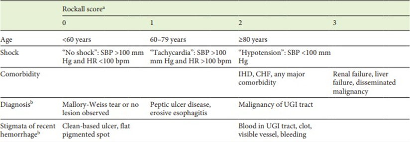
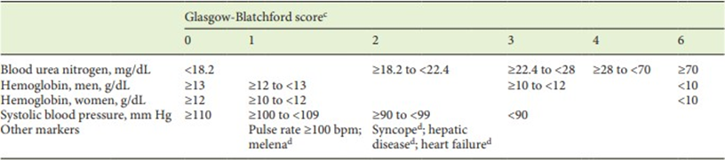
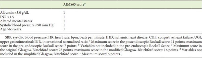
. Good system Glasgow Blatchford score (GBS) and also index shock is successful in predicting transfusion needs. The GBS system is more accurate than the shock index. However, because the GBS system is much more complex than index shock, index shock can become sign Which useful for with fast estimate need transfusion. However, GBS appears to have the best area under the curve values when this scoring system is used for predict need intervention endoscopy although No effective, followed by index shock. Mark area lower curve system AIMS65 seems to be the most low. Score GBS And AIMS65 can show mark Which high in certain individuals due to underlying gastrointestinal problems rather than the severity of MWS. Therefore, the GBS and AIMS65 systems may not be effective in the treatment of MWS caused by underlying gastrointestinal disorders. However, the GBS and AIMS65 scores are quite effective when used according to the algorithm in Figure 2 . 9
Table 1. Rockall, GBS and AIMS65 scores. 9
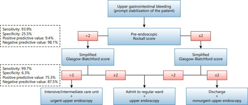
Picture 3. Algorithm Evaluation Risk Bleeding Conditions SCBA.9
Inspection Support
Laboratory tests include complete blood count, hemoglobin and hematocrit, and coagulation profile including bleeding time. (BT), time prothrombin (PT), time thromboplastin partial activated (aPTT), And amount platelets. Profile coagulation can used to assess the presence of severe thrombocytopenia and coagulopathy. If the platelet count is low with appropriate symptoms, the diagnosis of Mallory-Weiss syndrome should be considered, because chronic alcoholism as one of the predisposing factors for this syndrome can cause low platelet counts. 1,8
Renal function tests are also needed to recognize the presence of kidney failure by measuring blood urea nitrogen (BUN) and creatinine. If kidney failure occurs, it is most likely due to prerenal azotemia unless the patient has previous chronic kidney disease. Conditions of ischemia or myocardial infarction also need to be excluded by performing an electrocardiogram (ECG) and measuring cardiac enzymes. 1,8
Upper gastrointestinal endoscopy (URG) is the gold standard for diagnose tear Mallory Weiss in a way definitive, And treating simple active esophageal bleeding. During endoscopy, active bleeding, clots, or fibrin crusts may be found over the tear. In most case Mallory Weiss Syndrome can found linear tear single in part proximal curvature minor abdomen appropriate in below the cardia. SCBA endoscopy is also useful in finding other causes of bleeding including esophageal varices, gastric or duodenal ulcers. Most Mallory Weis tears are about one inch in length. 1.8
Radiological examination with barium is not recommended in this case because of its low diagnostic value and interference with endoscopic diagnosis. Meanwhile, angiography is indicated in actively bleeding tears if there is failure or inaccessibility of endoscopy to locate the bleeding tear and stop the bleeding. 1,8
Mallory Weiss syndrome must be differentiated from other causes of SCBA hemorrhage. It is important to differentiate between SMW and Boerhaave syndrome, Because both of them cause damage on esophagus. Syndrome Mallory-Weiss is a non-transmural tear of the esophagus, while Boerhaave syndrome is a transmural perforation of the esophagus. Esophageal rupture in Boerhaave syndrome is thought to result from a sudden increase in intraluminal pressure that happen during vomit, due to by coordination neuromuscular Which bad that cause failure muscle cricopharyngeus For relax. 6.7 Ulcer peptic ulcer is the cause most often from bleeding SCBA Which own description clinical characteristic and can be diagnosed with certainty by endoscopy. Esophageal varices, which are blood vessels winding widen in around esophagus part lower, part big as a complication hypertension portal Also can happen simultaneously with Mallory Weiss Syndrome. Besides That, malformation arteriovenous, neoplasm esophagus or stomach can also be a differential diagnosis of Mallory Weiss syndrome. 1,8
TREATMENT
Mallory-Weiss syndrome is mostly self-limiting and recurrence is rare, so initial management of the syndrome is aimed at stabilizing the patient's general condition. Immediate resuscitation of patients with active bleeding should be initiated on admission. Hemodynamic stability is assessed by checking the airway, breathing, and circulation (ABC protocol). Establishment of good central or peripheral intravenous (IV) access (usually 2 lines) along with fluid replacement therapy can be lifesaving in patients with massive bleeding. Packed red blood cell infusion is indicated if the hemoglobin level is less than 8 g/dL or if the patient presents with signs of shock or massive bleeding. 1,8
Decompression nasogastric use hose nasogastric can performed, especially in patients suspected of having esophageal varices, before gastric lavage. If electrolyte imbalance occurs, it must be corrected appropriately. Factor coagulation need optimized before continue with endoscopy. Most patients who are managed conservatively are usually hospitalized until hemostasis achieved And symptom resolved. 1.8 Maintenance conservative can be:
Therapy pharmacological
Proton pump inhibitors (PPIs) and H2 blockers are given to reduce gastric acidity, because increased acidity inhibits the recovery of the gastric and esophageal mucosa. Intravenous PPI drugs can be given to patients who will undergo an endoscopy. In addition, antiemetics such as promethazine and ondansetron can also be given to control nausea and vomiting. 1,8
Therapy endoscopy
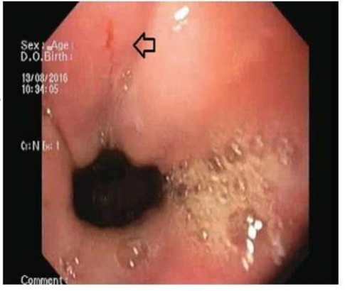
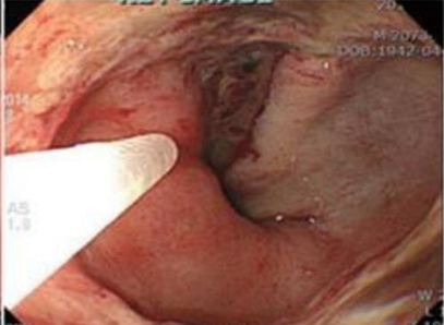
Although most patients can be managed with monitoring or conservative treatment, some cases require endoscopic or surgical treatment. For individuals with risk factors such as portal hypertension or coagulopathy, intensive care including endoscopic hemostasis recommended. On individual with bleeding active on Mallory-Weiss tear, inspection endoscopy very important For diagnosis and therapy (eg, arterial bleeding and diffuse bleeding). Stigmata on initial endoscopy (eg, visible vessels without bleeding and adherent clots) may not always require endoscopic therapy unless there have been recurrent bleeding episodes or associated coagulopathy. In such circumstances, endoscopic hemostasis is the recommended therapy. 1,8,10 As the endoscopic findings in Figure 4 , shown on picture adjacent left bleeding pulsatile still active from the esophago-cardial junction (arrow), while the right image shows the condition after hemostasis with cauterization together with thrombin 10,000 IU injection. 7
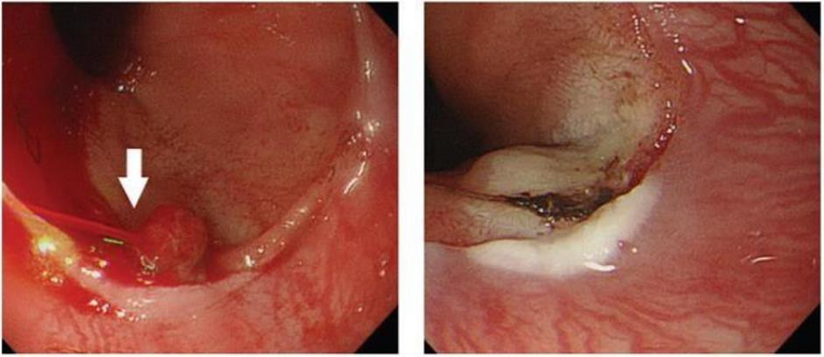
Picture 4. Endoscopic Findings Before And After Hemostasis. 7
Esophagogastroscopy is inspection choice in all SCBA bleeding cases. If the bleeding has stopped at the time of endoscopy, generally no further intervention is required. In situations with ongoing active or recurrent bleeding, there are therapeutic modalities endoscopy Which different. Injection local epinephrine For Stopping bleeding through vasoconstriction, multipolar electrocoagulation (MPEC), injection of sclerosing agents, contact heat treatment , argon plasma coagulation (APC), hemoclip implantation and band ligation are the most common endoscopic therapies for bleeding Mallory-Weiss tears. 1,8
endoscopic submucosal dissection (ESD) procedures , namely ESD traditional And endoscopic submucosal tunnel dissection (ESTD) as in Figure 6 . The esophageal lesion after iodine staining is shown in Figure 5A. While Figure 5B shows the submucosal tunnel created during ESTD. The artificial wound after ESTD is shown in Figure 5C, as well as mucosal laceration at the gastro-esophageal junction are shown in Figure 5D. 8 The standard ESD approach involves four steps, namely:
Marking limit lesion;
Injection submucosa For lift lesion;
Incision mucosa in around the lesion; And
Submucosal dissection, during which one or more submucosal tunnels are formed. The endoscopist then determines the ESD treatment based on the size of the lesion. 8
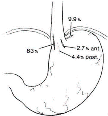
Picture 5. Location Most Frequent Tear Mallory- Weiss. 7
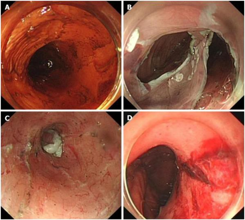
Picture 6. Appearance Tear Mallory Weiss on ESTD. 8
Therapy endoscopic injection
Therapy injection endoscopy use various type drug, Which most common is epinephrine (dilution 1:10,000 until 1:20,000). Injection therapy is choice maintenance Which simple And cheap. In terms of recurrent bleeding, length of hospital stay, and transfusion requirements, epinephrine injection therapy improves outcomes compared with supportive measures alone. Because epinephrine absorbed into the systemic circulation when used as an injection can cause ventricular tachycardia, this therapy should be avoided in patients with a history of coronary artery disease. 1,8,11
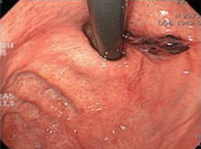
Picture 7. Therapy Endoscopic Injection . 7
Electrocoagulation endoscopy
Electrocoagulation allow implementation hot And pressure on bleeding lesions simultaneously. Coagulation is less effective in moist areas, like location bleeding Because hot with fast dissipated or lost by fluid. Proper device placement has proven difficult for lesions in the lesser curvature of the cardia. Multipolar electrocoagulation is significant increase hemostasis, reduce operational needs on patient with tear Mallory Weiss Which is bleeding, And Possible happen effect side minor. Coagulation repeat increase risk damage transmural And perforation Because relatively thin esophageal wall and loss of serosa at the site of tear. 1,8,11 Types therapy coagulation other Which available is argon plasma coagulation (APC) Where probe placed on distance from location of bleeding, And current electricity frequency tall, with rate flow gas argon that relatively low (1L/min), Then produce coagulation on the lesion bleeding. Absence contact between catheter And network produces wounds surface burn, reduce damage unwanted tissue and perforation.6,7
Implantation endoscopic hemoclip
Endoscopic hemoclip implantation is a simple therapy for non-fibrotic tissue bleeding lesions such as Mallory-Weiss tears and Dieulafoy ulcers. Because the bleeding location of Mallory-Weiss tears is at the gastro-esophageal junction , hemoclip placement can be quite difficult. Mechanism rotatable Which new found on catheter delivery allow adjustment hemoclip Which controlled And access easy to the gastro-esophageal junction . Dislodgement of hemoclips at the gastro-esophageal junction is common due to the large amplitude contractions in this anatomic region. In cases of deeper tear extension, such as with Boerhaave syndrome, placement of an endoclip can repair both edges of the tear and cover the perforated lesion. 1,8,11
Ligation band endoscopy
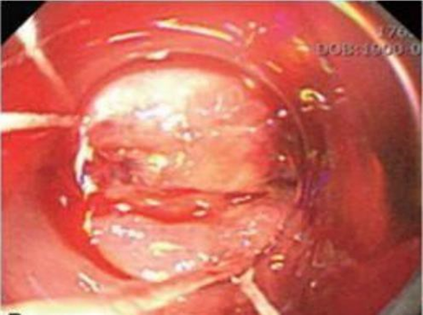
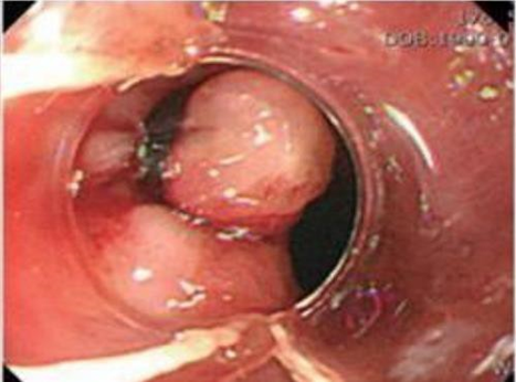
band ligation (EBL) has significant technological advantages compared to technique hemostatic other. Lesion seen clearly in a way tangential in EBL moment given pressure direct from the cover ligation transparent. When perforation esophagus happen, EBL very helpful For lesi bleeding in network non fibrotic. Closing which obviously makes EBL easier to operate by stopping the movement location bleeding And prevent movement from peristaltic including belching. The deeper portion of the visible vessel is ligated, providing permanent hemostasis and a strong ligation. Large Mallory-Weiss tears can be treated with a single band ligation . There was no difference in the effectiveness or safety of band ligation with epinephrine injection in a short, randomized prospective investigation of 34 individuals with actively bleeding lesions. Therapy thermal No recommended For tear Mallory Weiss associated with portal hypertension and gastric varices, so band ligation should be used . 1,8,11
Angiotherapy
Angiography with injection of a vasoconstrictor agent such as vasopressin or transcatheter embolization with gel foam for obliteration of the left gastric artery or superior mesenteric artery is considered when endoscopy is not available or fails. 1,8
Therapy surgery
Surgery is rarely necessary and is considered necessary after failure of endoscopic procedures or angiotherapy to stop the bleeding. In patients with severe upper gastrointestinal bleeding where a definitive diagnosis cannot be achieved before surgery, the surgeon should consider all choice. If No There is lesi duodenum or stomach, a wide gastrostomy is performed with laparotomy. At this time, blind gastrectomy is not recommended because of bleeding from the esophagus or cardia will missed And No treated. Reason bleeding will can be determined when the anterior wall of the stomach has been opened five to seven inches and gauze is applied. A Mallory-Weiss laceration in the proximal part of the stomach will be immediately visible and can be repaired with a running catgut suture . If the blood comes from esophagus, required level of exposure or visualization higher. Laparoscopic suturing Endoscopically guided rupture has shown excellent results. 1,8
Mallory-Weiss syndrome is a self-limiting disease in more than 90% of patients, so conservative treatment, including multiple transfusions, electrocoagulation, and compression with a Sengstaken-Blakemore tube is the preferred treatment, especially in patients with debilitated conditions. Sengstaken-Blakemore tube compression is the last resort in the treatment of bleeding Mallory-Weiss tears in patient Which weak. This is choice Which most No liked due to bleeding part big is artery And pressure on balloon No enough to overcome the pressure on the bleeding artery. Patients with cirrhosis have a very difficult time For treated And, let go from therapy, own prognosis Which bad. In a limited group of individuals, gastric prolapse into the esophagus may be the etiologic cause. 1,8
Conclusion
Mallory Weiss Syndrome (SMW) is a condition Which occurs due to a superficial longitudinal mucosal tear in the upper gastrointestinal tract, especially at the gastroesophageal junction. Although SMW is rare, the impact to health And condition patient can significant, especially on case with bleeding active or repetitive. As Wrong one cause bleeding channel digest part on (SCBA), SMW need careful diagnosis and proper management to achieve good results. In review anatomy, esophagus explained in a way comprehensive, describe structure And segments anatomy esophagus as well as blood supply And its nerves. Anatomy This become important in understand How tear Mallory Weiss can happen in intersection gastroesophageal and potentially cause bleeding. Identification of risk factors for SMW conditions is important in identifying groups of individuals at high risk and allowing effort prevention. Diagnosis SMW need comprehensive examination Which covering anamnesis Which careful, inspection physique and supporting examinations, including laboratory and endoscopy examinations which are very important important in to uphold diagnosis And evaluate level severity
bleeding.
SMW management depends on severity of bleeding and patient symptoms. Patients with active bleeding require hemodynamic resuscitation, stabilization, and endoscopic evaluation to stop the bleeding. Pharmacologic therapy with proton pump inhibitors (PPIs) helps in reducing gastric acidity. Endoscopy-based therapies such as epinephrine injection, electrocoagulation, And use method hemostasis other Also can performed according to initial endoscopic findings.
However, it should be noted that most cases of SMW can heal on their own and recurrence is rare. Therefore, the management of SMW depends on on evaluation And evaluation Which careful to symptom And severity of the patient's condition. In dealing with SMW conditions, the involvement of a multidisciplinary medical team is important to provide a correct diagnosis and effective management. Understanding deep about anatomy, factor risk, diagnosis, And governance SMW will help clinician give maintenance Which best for patients with this condition.
Ultimately, a comprehensive knowledge of Mallory-Weiss syndrome is key to identifying, diagnosing, and managing this condition. effective. With do approach Which holistic, including With appropriate and optimal monitoring and intervention for patients with SMW, complications can be prevented and patients can recover more quickly.
REFERENCE
Rawla P, Devasahayam J, Health S. Mallory-Weiss Syndrome . StatPearls Publishing; 2023. https://www.ncbi.nl m .nih.gov/books/NBK538190 /
Alban A, Alabbadi H, Almoqbell T, et a. The prevalence and risk factors for Mallory-Weiss syndrome: a systematic review and meta-analysis. Int J Community Med Public Heal . 2021;8(6):3096-3106. doi:10.1097/ICO.0000000000002150
Tortoise GJ, Derrickson BH. Principles of Anatomy and Physiology . 16th ed. John Wiley & Sons, Inc; 2020.
Chaudhry SR, Bordoni B. Anatomy, Thorax, Esophagus . StatPearls Publishing; 2022.
Chen W, Zhu XN, Wang J, Zhu LL, Gan T, Yang JL. Risk factors for mallory-weiss tears during endoscopic submucosal dissection of superficial esophageal neoplasms. World J Gastroenterol . 2019;25(34):5174-5184. doi:10.3748/wjg.v25.i34.5174
Hussain A, Kaler J, Tabrez E, Hussain S. The Presentation of Mallory-Weiss Syndrome Secondary to Underlying Pathologies. E.C. Gastroenterol Dig Syst . 2020;7(11):26-41.
Iqbal M, Lisfi I, Yusrawati. Mallory-Weiss Syndrome in Pregnancy. J Midwifery . 2022;7(2):98-112.
Hussein MS, Alfaraj FA, Alshabwi AD, et al. Evaluation and Management of Mallory–Weiss Syndrome: A Review. J Pharm Res Int . 2021;33(60A):788-794. doi:10.9734/jpri/2021/v33i60a34548
Cúrdia Gonçalves T, Barbosa M, Xavier S, et al. Optimizing the risk assessment in upper gastrointestinal bleeding: Comparison of 5 scores predicting 7 outcomes. GE Port J Gastroenterol . 2018;25(6):299-307. doi:10.1159/000486802
Radjabovich YF, Azimovich EE. Modern Treatment of Mallory-Weiss Syndrome. Int J Heal Syst Med Sci . 2023;2(4):27-33.
Lekshmi S, Soumya R, Prasobh G. A Review on Endoscopic Management of Mallory Weiss. World J Pharm Res . 2021;10(10):476-482.
Sugawa C, Benishek D, Walt A.J. Mallory-Weiss syndrome. A study of 224 patients. Am J Surg . 1983;145(1):30–3. https://doi.org/10.1016/0002- 9610(83)90162-9
Cucci M, Caputo F, Fraternity Orcion G, Roncallo A, Venture F. Transition of a Mallory-Weiss syndrome to a Boerhaave syndrome confirmed by anamnestic, necroscopic, and autopsy data: A case report. Medicine (Baltimore) . 2018;97(49):e13191
Jung DH, Ko BS, Kim YJ, Kim WY. Comparison of risk scores and shock index in hemodynamically stable patients presenting to the emergency department with nonvariceal upper gastrointestinal bleeding. Eur J Gastroenterol Hepatol . 2019;31(7):781–5. https://doi.org/10.1097/MEG.0000000000001422
Kim MS, Choi J, Shin WC. AIMS65 scoring system is comparable to Glasgow-Blatchford score or Rockall score for prediction of clinical outcomes for non-variceal upper gastrointestinal bleeding. BMC Gastroenterol . 2019;19(1):136. Available from: https://doi.org/10.1186/s12876-019-1051-8 him: 10.1186/s12876-019-1051- 8 https://doi.org/10.1097/MD.0000000000013191
Comments